Common Carotid Artery
The Common Vein Copyright 2009
Definition
The common carotid artery
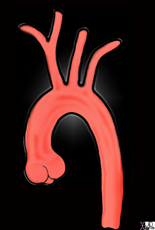
Origin of the Carotids |
|
This diagram shows the origin of the head neck and arm vessels from the arch of the aorta. The first vessel off the arch is the brachiocephalic or innominate artery and it branches into the right common carotid (RCC) and right subclavian artery. The second vessel is the left common carotid artery (LCC) and this is followed by the left subclavian artery. The RCC and LCC divide in the neck into the external and internal carotid vessels 47676b Courtesy Ashley Davidoff MD copyright 2010 |
To be edited
“The common carotid artery, of which there are two, are arteries which supply parts of the head and neck. The right common carotid artery arises from the brachiocephalic trunk, in back of the sternoclavicular joint. The left common carotid artery originates from the arch of the aorta. Both common carotids have cervical portions yet only the left has a thoracic portion. The thoracic portion of the left common carotid artery ascends from the arch of the aorta, through the superior mediastinum, and up to the level of the left sternoclavicular, where it becomes the cervical portion. In the front, the thoracic portion is separated from the manubrium sterni by the sternohyoideus and sternothyroideus, the front parts of the left pleura and left lung, the left brachiocephalic vein, and the remains of the thymus. In back it touches the trachea, esophagus, left recurrent nerve, and thoracic duct. On the right there is the brachiocephalic artery, as well as the trachea, the inferior thyroid veins, and the remains of the thymus, while on the left of the thoracic portion there is the left vagus nerve, as well as the phrenic nerve, the left pleura, and the left lung. The left subclavian artery is in back of and a little to the side of it. Both cervical portions of the right and left common carotid arteries run obliquely from the sternoclavicular articulatation up to a height level with the top of the thyroid cartilage. There, each divides into an external carotid artery and an internal carotid artery. The two common carotid arteries are separated from each other at their lower parts by a space containing the trachea, and at their upper parts by the thyroid gland, the larynx, and the pharynx. Each artery is encased in a sheath which also contains a few other veins and nerves, though each vessel as a separate compartment within this encasement. In back, the common carotid arteries are separated from the trasnverse process of the cervical porcesses by the longus colli and longus capitis. Near the center of the body they are close to the esophagus, trachea, thyroid gland, larynx, phrarynx, inferior thyroid artery, and recurrent nerve. Farther from the center they are close to the internal jugular vein and vagus nerve. The arteries are crossed at their terminations by the superior thyroid vein, a little below the cricoid cartilage by the middle throid vein, and a little above the clavicle by the anterior jugular vein. There is a triangle, called the carotid triangle, formed by the sternocleidomastoideus in back, the stylohyoideus and diagastricus above, and the omohyoideus below, through which the common carotid arteries can be seen. These portions of the arteries are crossed by the sternocleidomastoid branch of the superior thyroid artery and the superior and middle thyroid veins. The arteries are also crossed in back by the right recurrent nerve and the left internal jugular vein often overlaps them. In the lower part of the neck, the common carotid arteries are covered by intergument, subcutaneous fascia, platysma, deep cervical fascia, the sternocleidomastoideus, the sternohyideus, the sternothyroideus, and the omohyoideus. In the upper part of the neck, however, they are only covered by intergument, superficial fascia platysma, deep cervical fascia, and the medial margin of the sternocleidomastoideus. A small oval body called the carotid body is located near the common carotid arteries. It is 2-5 mm in diamter, composed of epithelioid cells, nerve fibers, and a fibrous capsule. The carotid body senses pH, carbon dioxide, and oxygen concentrations in the blood and is a part of the homeostatic control system. There is also a dilatation near the bifurcation of the common carotid arteries and the internal carotid arteries, called the carotid sinus, which contains nerves to regulate blood pressure. The right common carotid artery, is twelve percent of cases, originates above the top level of the sternoclavicular articulation. It can also arise directly from the arch of the aorta or from a trunk shared with the left common carotid artery. The left common carotid artery’s point of origin is different in some patients. Occassionally the common carotid arteries do not give off external or internal carotids, and sometimes the common carotid itself is absent and the external and internal carotids arise directly from the arch of the aorta. In some instances, the common carotid arteries give off a superior thyroid branch, a laryngeal branch, the ascending pharyngeal artery, the inferior thyroid artery, or the vertebral artery.”
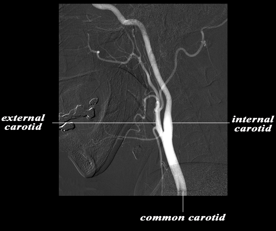
The Common Carotid and Terminal Branches |
|
The angiogram demonstrates the carotid system in the lateral projection. The common carotid artery bifurcates in the base of the neck into the external and internal carotid artery. Basically the internal carotid artery supplies the brain and the external supplies the structures of the face and muscles and skin of the cranium. The carotid system supplies the brain from the internal carotid – a branch of the common carotid which arises from the aorta. Courtesy Ram Chavalli MD 49400bc01.8s |

Common Carotid in Axial projection and Neighbours in the Neck |
|
The axial CT demonstrates the central position of the thyroid in the neck and its relations. Anteriorly a group of muscles called the strap muscles (tan) separate the thr thyroid gland from the skin. The thyroid surrounds the trachea (teal) on its anterior and lateral borders. The esophagus (darker pink) lies posterior to the trachea and medial to the posterior brder of the lobes of the thyroid. The common carotid arteries lie posterior to each of the lobes, while the internal jugular veins lie lateral to the lobes. The prevertebral muscles (light brown) and vertebral arteries (bright red) form posterior relations of the thyroid. Courtesy Ashley Davidoff MD copyright 2010 all rights reserved 30491c02b02L.9s |
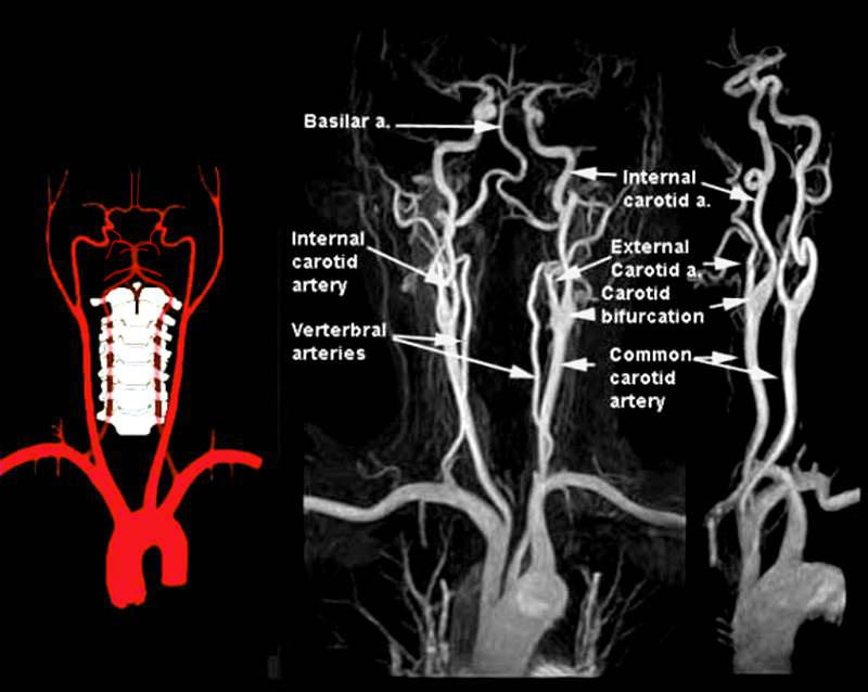
The Cephalic Arteries |
|
The diagram shows the main branches of the blood supply to the brain which includes the carotid and vertebro-basilar systems. The carotid system supplies the brain from the internal carotid – a branch of the common carotid which arises from the aorta. On the right side the common carotid arises from the brachiocephalic artery and on the left it arises as the common carotid as the second branch off the aortic arch It enters the cranial cavity through the cavernous sinus and gives rise to two main branches; the anterior cerebral and the middle cerebral The vertebral arteries usually arise from the subclavian arteries and then travel in the vertebral foramina in the cervical spine. The basilar artery is formed by the two vertebral arteries and travel as a single artery over the upper medulla and the entire pons. Its terminal division is into the right and left posterior cerebral arteries. Its first branch after this division is the posterior communicating artery. It continues around the pons and midbrain in the ambient and quadrigeminal cisterns and then ascends above the tentorium to end in the occipital lobe. Each of the vessels contributes to the circle of Willis through communicating arteries. The vertebro-basilar system provides the posterior communicating arteries bilaterally and the carotid system provides the anterior communicating arteries via the middle cerebral artery. The PICA or posterior inferior cerebellar artery arises from the vertebral arteries and the anterior inferior cerebellar artery arises from the basilar artery. The superior cerebellar artery arises from the tip of the basilar artery just before it terminates in the posterior cerebral arteries Courtesy Philips Medical Systems 92492.8 |
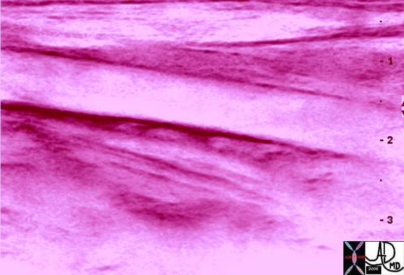
The Common Carotid Artery Normal – Ultrasound |
|
The artistic rendering overlays the ultrasound a normal common carotid artery in longitudinal view in the the neck 46493b02 Courtesy Ashley Davidoff MD Copyright 2010 |
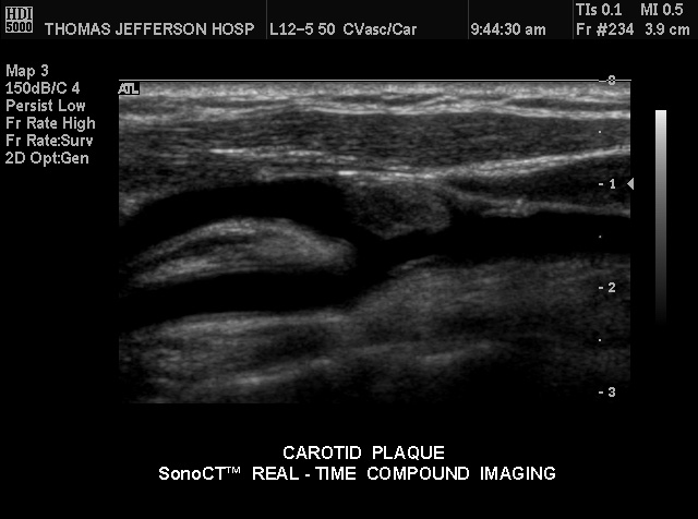
Plaque of the Distal Common Carotid |
| Patient with plaque at the distal end of the common carotid artery just prior to the carotid bifurcation demonstrated by conventional gray scale US technique .
Courtesy Philips Medical Systems 33271 |
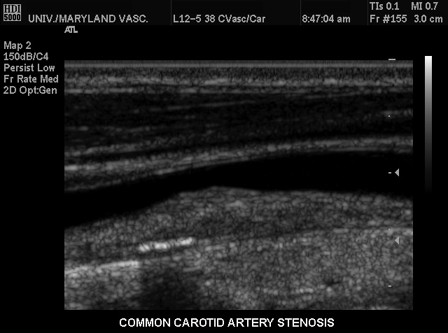
Moderate Stenosis Atherosclerosis |
| This conventional gray scale US of the neck shows a common carotid artery heaped up plaque on the far wall causing stenosis of moderate degree. Note also the linear area of strong echogenicity on the far wall associated with shadowing consistent with a calcification in the wall. These findings are characteristic of atherosclerotic plaque.
Courtesy Philips Medical Systems 33289 |
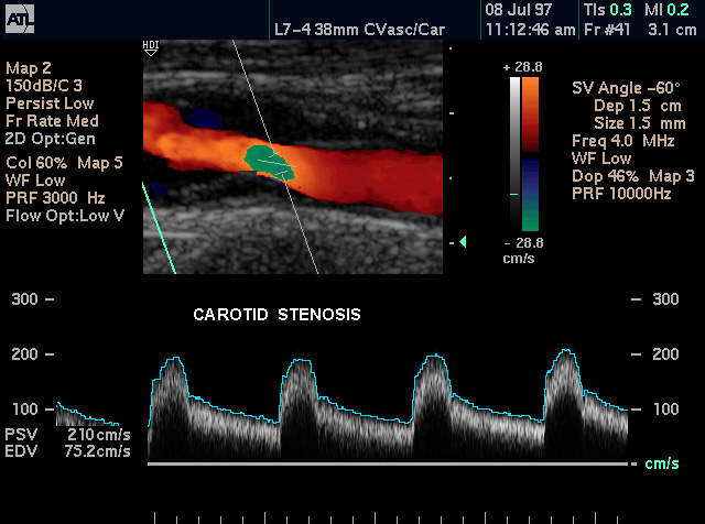
Color Flow Doppler of Mild Carotid Stenosis |
| Patient with plaque in the common carotid artery causing carotid stenosis demonstrated by color dopppler US technique .
Courtesy Philips Medical Systems 33272 |
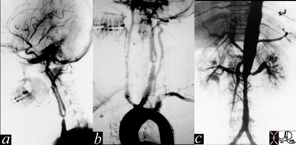
Takayasu’s Arteritis |
| The series of images are from the angiogram of a 14 year old female who presented with seizures and an elevated blood pressure. Images a and b show multiple stenoses within the carotids best seen at the level of the bifurcation into external and internal arteries. In addition in b, the aortic arch shows non critical narrowing just after the origin of the left common carotid vessel. Note that the right subclavian artery is not seen and presumably is accluded at its origin. The abdominal angiogram shows a significant narrowing of the left renal artery with post stenotic dilitation, and stenotic disease in the infrarenal abdominal aorta. The multicentric nature of the disease in a young female is athognomonic of Takayasu’s arteritis.
35155c Courtesy of Laura Feldman MD. |
End of Life No Intracerebral Flow |
|
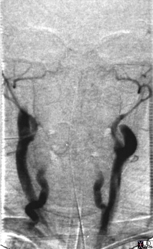
This image represents the sad ending of life for this individual. The extracranial vessels are all occluded and there is no intarcerebral flow or perfusion. These findings are compatible with brain death. 22772 Courtesy Ashley Davidoff MD |
