Copyright 2010
Definition
The substantia nigra is a region of gray matter in the tegmentum of the midbrain situated posterior to the cerebral peduncles and and anterior to the red nuclli. The dopaminergic neurons that make it up contain melanin giving it a black appearance – and hence its name.
Functionally it is linked to the basal ganglia and also movement and more specifically eye movement. It is also responsible for dopamine production, which has been linked to reward seeking behavior as well as addiction.
Parkinson’s disease is a failure of dopaminergic neurons of the substantia nigra
|
Substantia Nigra (black bands) |
|
The anterior border of the midbrain incorporates the cerbral peduncles(green), and the substantia nigra (black) – just posterior to the peduncles. Between the substantia nigra and the aqueduct is an area of the midbrain called the tegmentum (floor of the midbrain) The posterior end of the midbrain is bordered by the colliculi (purple) in the tectum (roof) of the midbrain. Courtesy Ashley Davidoff MD copyright 2010 all rights reserved 94074b09b05b.83s |
|
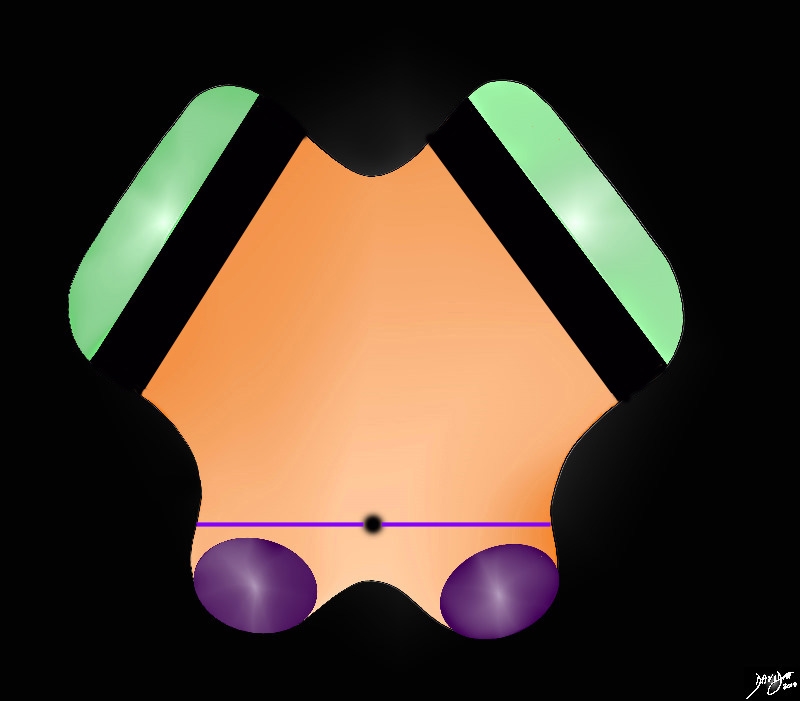
Cerebral Peduncles in Axial Projection |
|
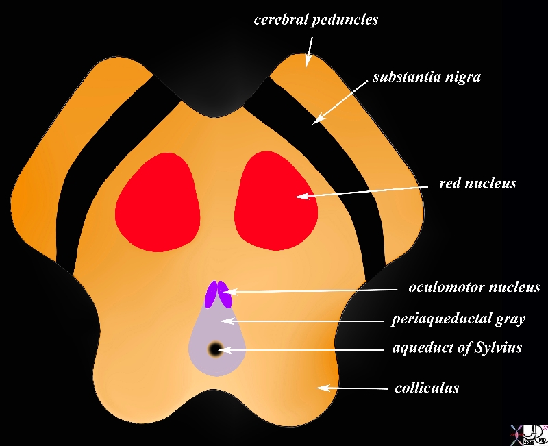
The midbrain in transverse plane illustrates the component structures, with the image reminiscent of the face of a baby pig.. Anteriorly the cerebral peduncles are followed by the substantia nigra, and the collilculi The anterior border of the midbrain incorporates the cerebral peduncles, and the substantia nigra (black – just posterior to the peduncles). Between the substantia nigra and the aqueduct is an area of the midbrain called the tegmentum (floor of the midbrain) Within the tegmentum other structures include red nuclii, oculomotor nuclii, periaquaductal gray, and the aqueduct of Sylvius which is border forming between the tegmentum anteriorly and the tectum (roof) posteriorly. The posterior end of the midbrain is bordered by the colliculi in the tectum. Courtesy Ashley Davidoff MD copyright 2010 all rights reserved 94074b08a06bL.9s |
|
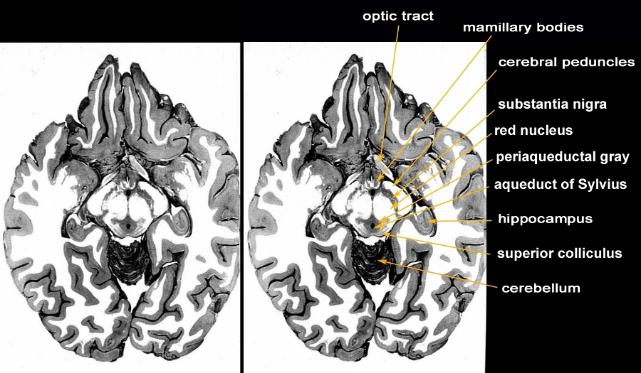
Axial Anatomic Specimen through the Midbrain Periaqueductal Gray Around the Aqueduct of Sylvius |
|
The anatomic section is an axial slice through the brain showing the structures of the midbrain. The most anterior structures of the midbrain are the cerebral peduncles followed by the substantia nigra, red nucleus, periaquaductal gray (PAG or central gray), aqueduct of Sylvius and finally the superior colliculus. Structures that are related to the midbrain anteriorly are the mamillary bodies and the optic tract. The hippocampus is positioned posterolaterally and the superior aspect of the cerebellum is seen posteriorly. Courtesy Department of Anatomy and Neurobiology at Boston University School of Medicine Dr. Jennifer Luebke , and Dr. Douglas Rosene 97140b03c.9 |
|
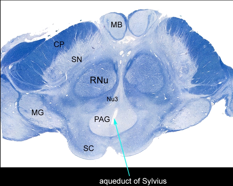
Whole Mount of the Midbrain in Axial Projection |
|
The mounted and stained midbrain in transverse plane illustrates the component structures. The anterior border of the midbrain incorporates the cerebral peduncles (CP), and the substantia nigra (SN). Between the substantia nigra and the aqueduct (teal arrow) is an area of the midbrain called the tegmentum (floor of the midbrain) Within the tegmentum other structures include red nuclii (RNu), oculomotor nucleus (Nu3), periaquaductal gray (PAG), and the aqueduct of Sylvius which is border forming between the tegmentum anteriorly and the tectum (roof) posteriorly. The posterior end of the midbrain is bordered by the colliculi in the tectum. Note also the paired mamillary bodies anteriorly (MB) and the medial geniculate body (MG) posterolaterally Courtesy Department of Anatomy and Neurobiology at Boston University School of Medicine Dr. Jennifer Luebke , and Dr. Douglas Rosene 98483L.8 |
|
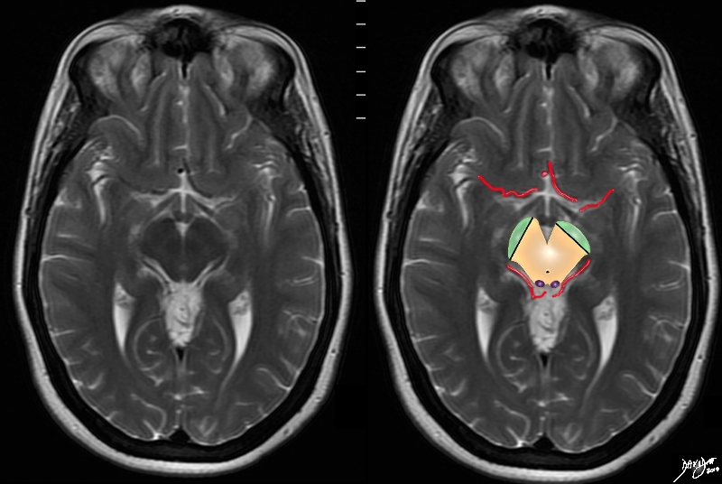
The Cerebral arteries Characteristically Surround the Midbrain |
|
The MRI T2 weighted series now focuses on another very characteristic feature of the midbrain in that it is surrounded by the anterior and posterior cerebral circulations, and in this case portions of the arteries are seen with parts of the middle cerebral seen anteriorly and parts of the posterior cerebral seen posteriorly Davidoff art Courteys Ashley Davidoff MD copyright 2010 all rights reserved 94081.4kc02.8s |
Sagittal Plane
|
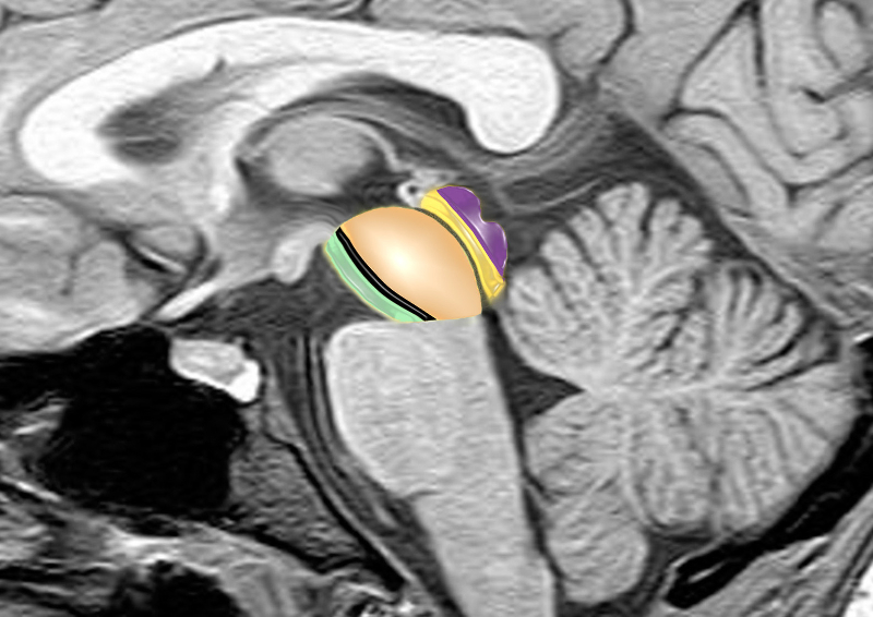
Basic Divisions of the Midbrain Tegmentum and Tectum Cerebral Peduncles, Cerebral Crura and Colliculli |
|
The midbrain consists of a larger anterior portion consisting of the cerebral peduncles, (green) the substantia nigra (black) and the tegmentum (light orange) that extends to the aqueduct (thin gray line) The posterior portion called tectum (orange) contains the colliculi (purple) which are the most posterior portions of the midbrain. brain anatomy neuroanatomy midbrain tegmentum colliculi tectum cerebral peduncle aqueduct of Sylvius conceptual diagram MRI principles Image provided by Philips Medical Systems Enhanced by Davidoff art Courtesy Ashley Davidoff MD 92141.3kb01ba02b02.8s |