
Posterior Cerebral Artery
The Common Vein Copyright 2010
Introduction
Origin
It is the terminal branch of the basilar artery, originating just below the foramen ovale of Pacchioni.
Branches To and From the Circle of Willis |
|
This cerebral circulation is centered on the circle of Willis which has two basic systems that feed it; the carotid system (in this case salmon pink) and the vertebro-basilar system (brighter pink). They both feed into the circle of Willis (bright red) via communicating branches. The middle cerebral artery is the vessel that feeds into the COW by providing the anterior communicating artery and the posterior cerebral artery connects via the posterior communicating artery. Conceptually as depicted in this diagram the circle of Willis is the centre of the cerebral circulation and from it blood flows into the anterior (bright green), middle (darker green) and posterior cerebral (maroon) regions. Image Courtesy Ashley Davidoff MD Copyright 2010 97194b16b.8s |
|
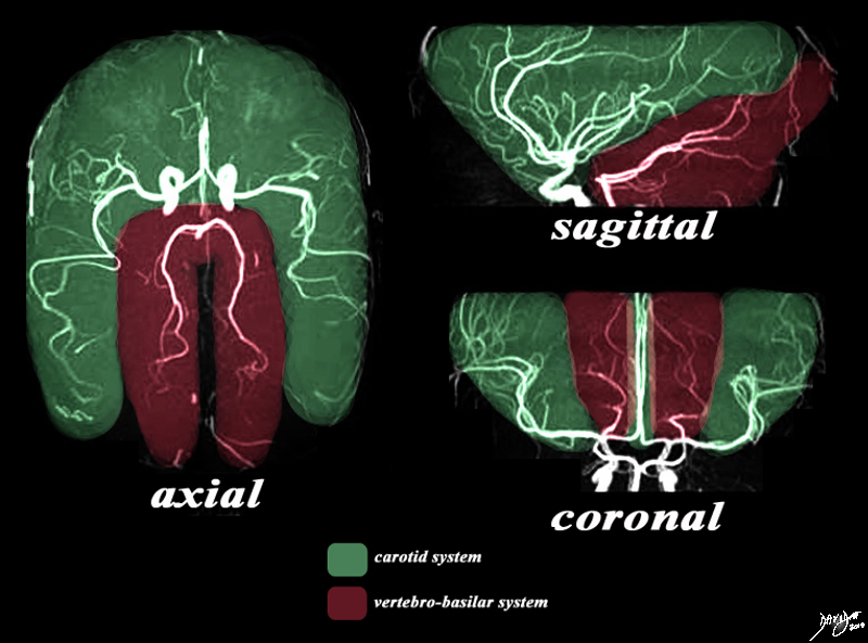
The Two Systems – Carotid and Vertebrobasilar |
|
There are two systems that supply the brain with blood. The MRA depicted in the axial, sagittal, and coronal views depict the general areas covered by the two systems. The carotid system gives rise to the anterior and middle cerebral arteries which cover the green area. The vertebro-basilar system, mostly through the posterior cerebral artery and cerebellar arteries, supply the maroon portion. Image Courtesy Philips Medical Systems Artistic rendering Davidoff MD Copyright 2010 92479c03.8s |
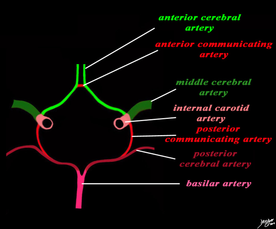
Structures of the Circle of Willis |
|
The diagram shows the main branches of the blood supply to the brain which includes the carotid and vertebro-basilar systems. These are the vessels that particpate in the formationofthe circle of Willis The carotid system supplies the brain from the internal carotid (salmon pink) – a branch of the common carotid which arises from the aorta. Its terminal bifuracation into the middle cerebral (dark green) and anterior green (bright green) are shown. The anterior communicating artery runs between the two anterior cerebrals (bright red) The basilar artery (pink) is formed by the two vertebral arteries and travel as a single artery over the upper medulla and the entire pons. Its terminal branch is the posterior cerebral artery (maroon). Each of the vessels contributes to the circle of Willis through communicating arteries. The vertebro-basilar system provides the posterior communicating arteries bilaterally and the carotid system provides the anterior communicating arteries via the anterior cerebral artery. code brain Courtesy Ashley Davidoff MD Copyright 2010 All rights reserved 97194b13g04L01.91s |
|
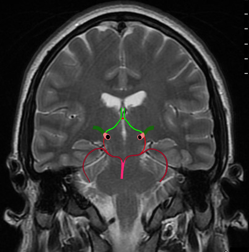
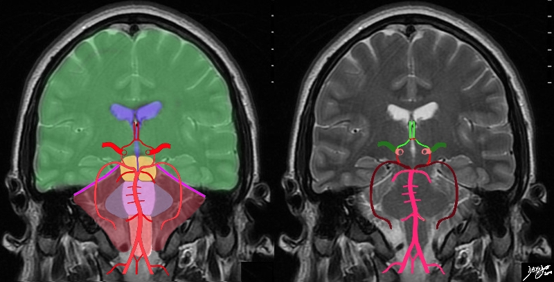
The Circle of Willis Overlaid on a Coronal View of the Brain |
|
The diagram shows the main branches of the blood supply to the brain including the circle of Willis overlaid on coronal MRI image to portray the approximate position of the vessels in the brain. The carotid system supplies the brain from the internal carotid we demonstrate its terminal bifurcation into middle cerebral (dark green) and anterior cerebral (bright green). The anterior communicating artery runs between the two anterior cerebrals (bright red) The basilar artery (pink) is formed by the two vertebral arteries and it travels as a single artery over the upper medulla and the pons. Its terminal branch is the posterior cerebral artery (maroon). Each of the carotid and vertebro-basilar systems contributes to the circle of Willis through communicating arteries. The vertebro-basilar system provides the posterior communicating arteries bilaterally from the posterior cerebral and the carotid system provides the anterior communicating arteries via the anterior cerebral arteries. Courtesy Ashley Davidoff MD Copyright 2010 All rights reserved 89721c06b.8sg04.8s |
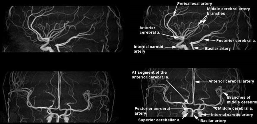
Map of the Cerebral Circulation |
|
The MRA in sagittal (upper images) and coronal projections (lower images) provide a map of the first and second order branches of the anterior and middle cerebral vessels. Image Courtesy of Philips Medical Systems 92484b.9s |
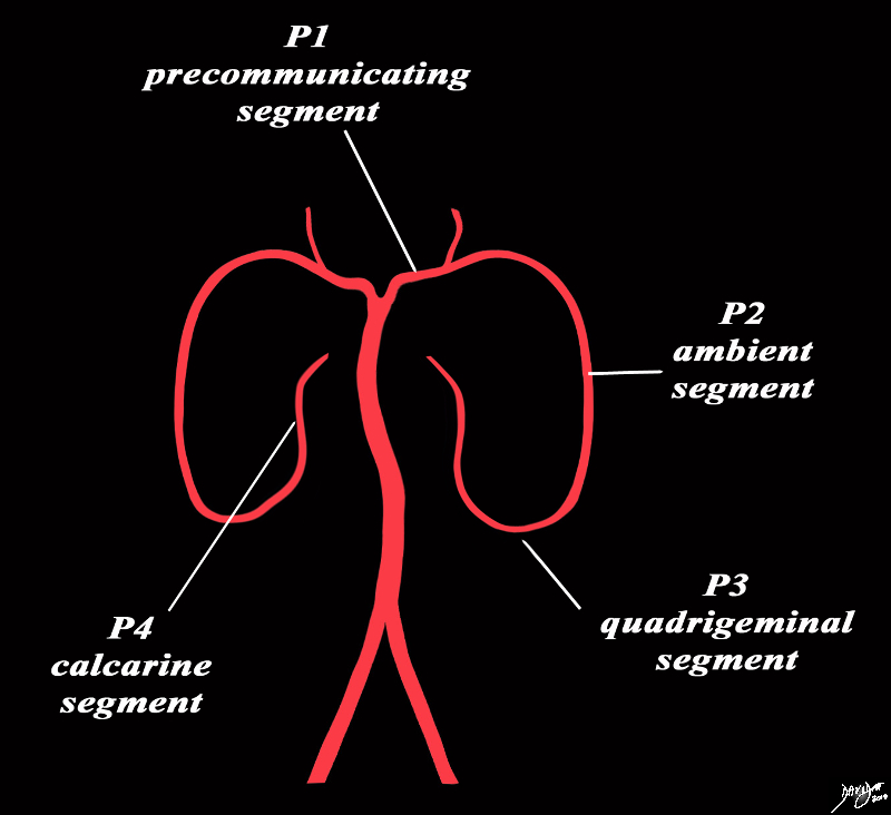
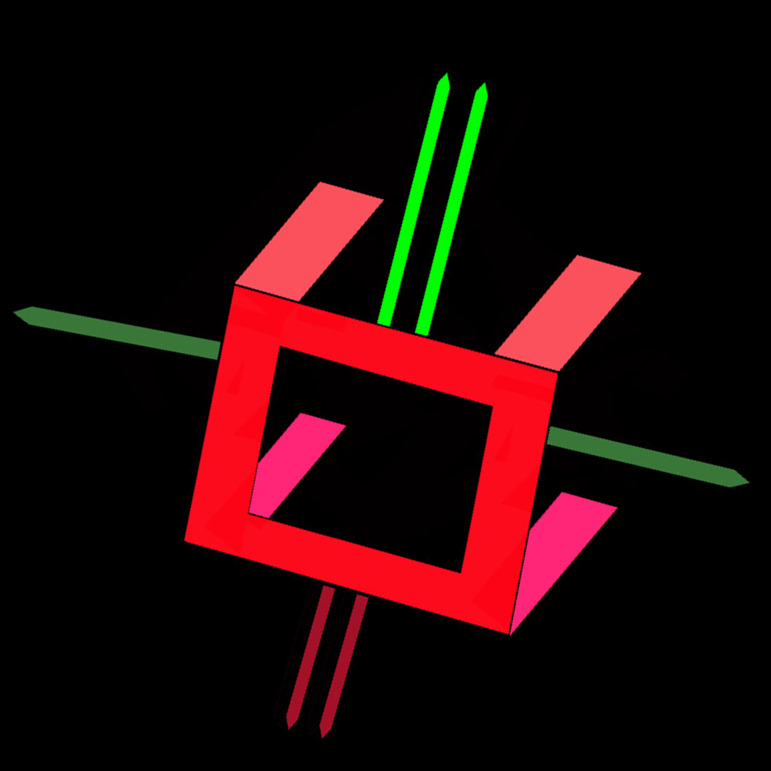
The Posterior Cerebral Artery Frontal View |
|
The diagram shows divisions of the posterior cerebral artery. The origin of the posterior communicating artery defines the border between the P1 segment (precommunicating segment) and the P2 segment (ambient segment) which proceeds laerally around the peduncles over the superior border of the pons in the region of the ambient cistern. The P3 segment is a short segment which is called the quadrigeminal segment as it is in close proximity to the quadrigemminal plate and within the quadrigeminal cistern. The P4 segment proceeds posteriorly and above the tentorium to the region of the occipital lobe and calcarine fissure. Courtesy Ashley Davidoff MD copyright 2010 all rights reserved 97194b06b03g.8s |
Course
It has a pre-communicant segment – P1 segment – located between the basilar artery and the anastomosis with the posterior communicating artery. It contours the midbrain in the lateral part.
P2 segment (ambient segment), runs over the basal vein of Rosenthal, being in relation with the hippocampal fissure. It contours the lateral superior colliculus of the midbrain.
The P3 segment starts at the superior colliculus. It enters the inner surface of the temporal and occipital lobes, ending at the calcarine fissure. The terminal branch is the calcarine artery.
|
The vertebral Arteries Part of the vertobasilar System |
|
The diagram shows the main branches of the blood supply to the brain including the circle of Willis overlaid on coronal MRI image to portray the approximate position of the vessels in the brain. The image on the right shows the combined system all in red, and the image on the right shows the derivation from the vertebrobasilar and carotid systems The carotid system supplies the brain from the internal carotid (salmon pink). We demonstrate its terminal bifurcation into middle cerebral (dark green) and anterior cerebral (bright green). The anterior communicating artery runs between the two anterior cerebrals (bright red) The basilar artery (pink) is formed by the two vertebral arteries and it travels as a single artery over the upper medulla and the pons. Its terminal branch is the posterior cerebral artery (maroon). The first branch off the posterior cerebrals is the posterior communicating which joins the middle cerebral to complete the circle of Willis Each of the carotid and vertebro-basilar systems contributes to the circle of Willis through communicating arteries. The vertebro-basilar system provides the posterior communicating arteries bilaterally from the posterior cerebral and the carotid system provides the anterior communicating arteries via the anterior cerebral arteries. Courtesy Ashley Davidoff MD Copyright 2010 All rights reserved 89721c06b.8sg05.8s |
|
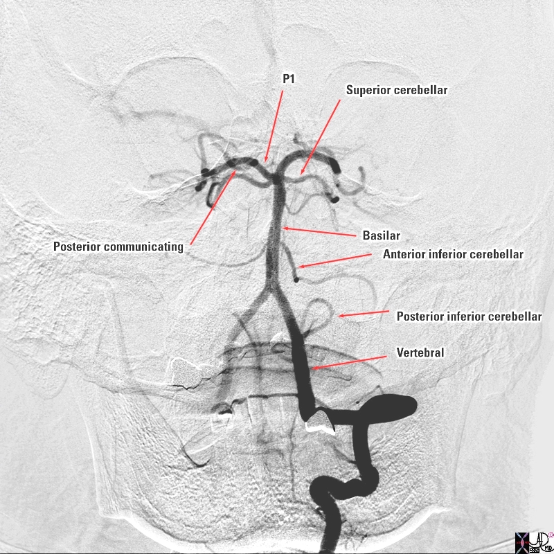
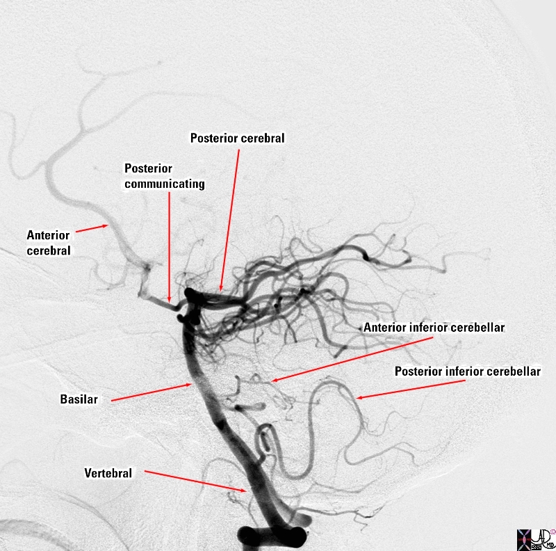
The Vertebral Artery and Major Branches Selective Angiogram in the A-P Projection |
|
The selective of the left vertebral artery in the A-P projection reveals the major intracranial branches; The posterior inferior cerebellar artery (PICA) which supplies the inferior aspect of the cerebellum The two vertebral arteries join to form the basilar artery which runs on the anterior surface of the pons. Its first branch is the; anterior inferior cerebellar artery ( AICA) which supplies the middle portion of the cerebellum and then the basilar artery gives off the superior cerebellar artery which supplies the superior aspect of the cerebellum The basilar artery finally terminates in the two posterior cerebral arteries (PCAs) which give off the posterior communicating arteries which connect with the circle of Willis and feed the inferior portion of the temporal lobes and the occipital lobes. Courtesy Elisa Flower MD and Alex Norbash MD Copyright 2010 97629b.8s |
|
The Slective Angiogram in the Lateral Projection |
|
The selective angiogram of the vertebral artery in the lateral projection reveals the major intracranial branches; The posterior inferior cerebellar artery (PICA) which supplies the inferior aspect of the cerebellum The two vertebral arteries join to form the basilar artery which runs on the anterior surface of the pons. Its first branch is the; anterior inferior cerebellar artery ( AICA) which supplies the middle portion of the cerebellum and then the basilar artery gives off the superior cerebellar artery which supplies the superior aspect of the cerebellum (not easily distinguished in this view The basilar artery finally terminates in the two posterior cerebral arteries (PCAs) which give off the posterior communicating arteries which connect with the circle of Willis. The PCA feeds the inferior portion of the temporal lobes and the occipital lobes. The anterior cerebral artery is seen filling from the circle of Willis Courtesy Elisa Flower MD and Alex Norbash MD Copyright 2010 97630b02.8s |

Vertebral Circulation – A-P Projection |
| The A-P projection of the right vertebral injection shows cross filling into the left vertebral, the basilar artery and the posterior cerebral artery. The posterior inferior cerebellar artery (PICA) is the first branch off the basilar artery but its origin off the basilar is slightly more difficult to appreciate. The origin and course of the the superior cerebellar artery is is easily seen and recognized as it arises just inferior to the larger posterior cerebral arteries which are the terminal branches of the basilar artery. In the A-P projection they assume a vertical course as they make their way to the occipital lobes.
49418 Courtesy Ram Chavalli |
Branches
mesencephalic circumflex arteries
paramedian thalamic perforating artery
thalamo-geniculate arteries
posteromedial choroidal artery
temporal lobe arteries
occipital lobe arteries
Territory
Superficial branches:
Inferomedial temporal cortex
Medial face of the occipital lobe
Posterior portion of the cingulate gyrus
Splenium of the corpus callosum
Deep branches:
Superior and posterior regions of thalamus
Subthalamic region
Cerebral peduncles
Choroid plexus
Applied Biology
Disease
Superficial unilateral infarction: contralateral homonymous hemianopia; alexia (dominant lobe infarcts)
Bilateral superficial infarction: cortical blindness
Profound unilateral infarction (thalamic lesion): contralateral hemi-hyposthesia of all senses
Profound bilateral infarction: amnesic syndrome, Korsakov-type
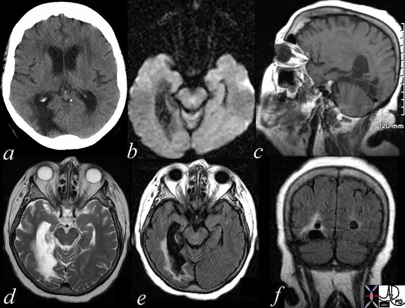
Chronic Infarct Posterior Cerebral Circulation |
|
The patient is a 74 year old female with a history of ataxia, incontinence, and memory loss. The MRI shows features of a chronic infarction in the right posterior cerebral artery territory affecting the posteromedial aspect of right temporal lobe and medial and inferior aspect of the right occipital lobe. There is evidence of gliosis with volume loss, atrophy, and encephalomalacia resulting in ex vacuo changes with dilatation of temporal and occipital horns of the lateral ventricles. Image a is a CTscan showing encephalomalacia in the right posterior temporal region and ex vacuo change with ventricular dilatation. Image b is a DWI scan showing no acute changes. Image c is a T1 weighted sagittal view showing no acute hemorrhage. Image d is a T2 weighted study showing compound high intensity changes in the region of the occipital horn but the distinction between disease and dilated ventricle cannot be made because of blooming and isointensity of the ventrciles and the diseases cerebral tissue. Image e is a FLAIR image that turns the CSF black and enables the identification of the high intensity abnormal brain tissue that is smaller than expected after reviewing the T2 weighted image. This case reveals the importance of the FLAIR image. Image f is a coronal FLAIR sequence also showing that the actual dimension of diseased tissue is relatively small. The findings are consistent with an old chronic infarction. Courtesy Ashley Davidoff MD Copyright 2010 All rights reserved 71419c01.800 |
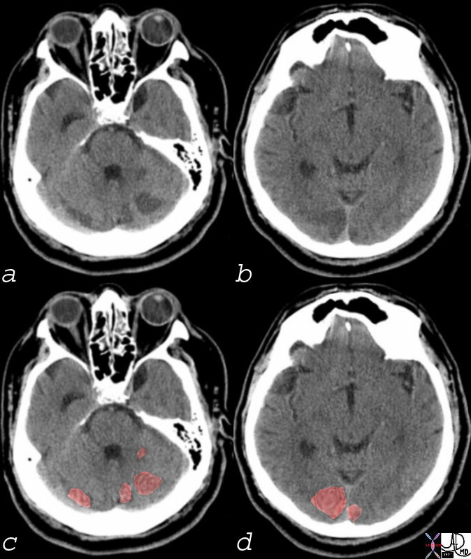
Multicentric Infarcts in the Cerebellum and Occipital Lobes |
|
Multicentric low density regions in both posterior cerebellar hemispheres and the occipital lobes bilaterally seen in (a and b) correspondingly overlaid in light red in c and d, are consistent with non hemorrhagic infarcts. Embolic disease is most likely Courtesy Ashley Davidoff MD 74923c01 |