
Middle Cerebral Artery
Copyright 2010
Introduction
Origin
The MCA is the largest of all cerebral arteries and it is a terminal branch of the internal carotid artery. It actually seems to continue it in terms of direction and caliber. This anatomical feature explains the higher greater frequency of ischemic strokes in this territory. Its vast territory stretches over the entire outer surface of the hemispheres.
Supplies the almost the entire lateral surface of the hemisphere; except for the small superior inch of the frontal and parietal lobe which is supplied by the frontal(anterior cerebral artery), and the inferior part of the temporal lobe.
to the cortical areas involved in speech, language and swallowing.
left middle cerebral artery provides Broca’s area, Wernicke’s area, Heschl’s gyrus, and the angular gyrus
most of the blood supply to the corpus striatum = basal ganglia and internal capsule.
A rupture of the lenticulo-striate artery results in bleeding usually in the region of the internal capsule
lenticulostriate arteries have relatively thin walls more vulnerable to hemorrhage
Course:
Its course has four segments:
M1 segment, or sphenoid portion: parallel to the lesser wing of sphenoid that gives branches to the basal ganglia. This segment provides the brain with numerous (lateral lenticulostriate) arteries that irrigate the basal ganglia.
M2 segment, also named the insular portion, when it courses in this lobe.
M3 segment, or opercular segment, which corresponds to the portion that follows the curve of the operculum over the surface of the insula.
M4 segment, or the terminal/cortical part, which is located at the bottom of the lateral fissure
Branches
The M1 segment provides the brain with numerous (lateral lenticulostriate) arteries that irrigate the basal ganglia.
The M2 segment gives a lateral frontobasilar branch, just before it emerges in the insula. Soon when it enters it, it provides collaterals to the temporal lobe – the anterior temporal artery, middle temporal artery and posterior temporal artery.
Later, in the M2/M3 segments it gives branches to to the central sulcus, precentral sulcus, postcentral sulcus, as well as anterior and posterior parietal arteries and finally an artery to the angular gyrus.
Branches To and From the Circle of Willis |
|
This cerebral circulation is centered on the circle of Willis which has two basic systems that feed it; the carotid system (in this case salmon pink) and the vertebro-basilar system (brighter pink). They both feed into the circle of Willis (bright red) via communicating branches. The middle cerebral artery is the vessel that feeds into the COW by providing the anterior communicating artery and the posterior cerebral artery connects via the posterior communicating artery. Conceptually as depicted in this diagram the circle of Willis is the centre of the cerebral circulation and from it blood flows into the anterior (bright green), middle (darker green) and posterior cerebral (maroon) regions. Image Courtesy Ashley Davidoff MD Copyright 2010 97194b16b.8s |
|
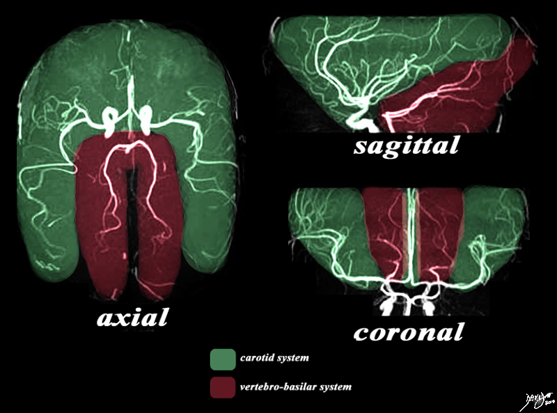
The Two Systems – Carotid and Vertebrobasilar |
|
There are two systems that supply the brain with blood. The MRA depicted in the axial, sagittal, and coronal views depict the general areas covered by the two systems. The carotid system gives rise to the anterior and middle cerebral arteries which cover the green area. The vertebro-basilar system, mostly through the posterior cerebral artery and cerebellar arteries, supply the maroon portion. Image Courtesy Philips Medical Systems Artistic rendering Davidoff MD Copyright 2010 92479c03.8s |
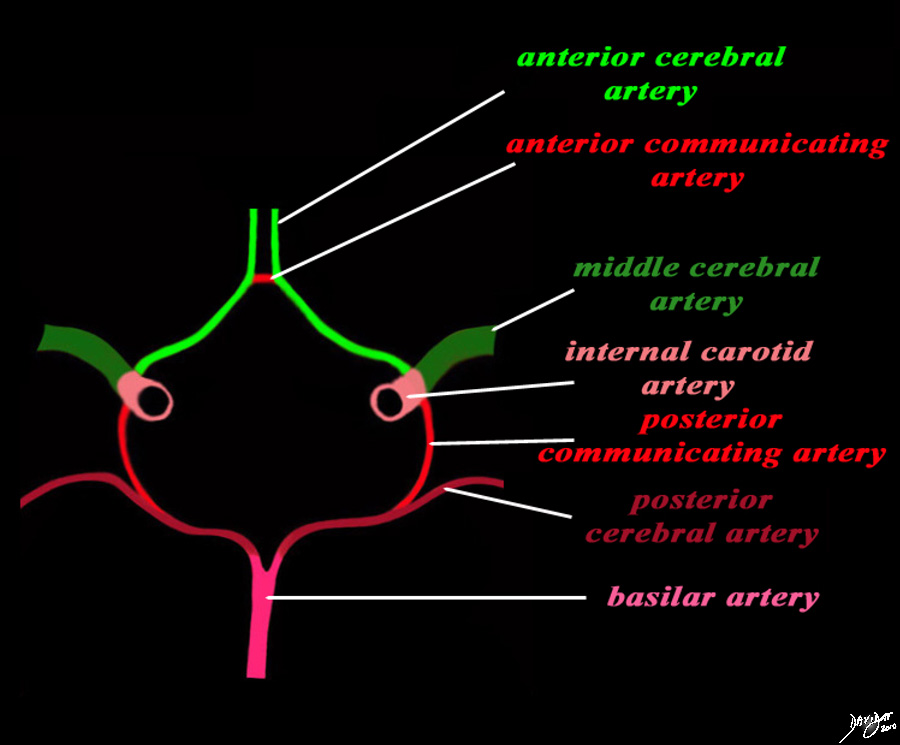
Structures of the Circle of Willis |
|
The diagram shows the main branches of the blood supply to the brain which includes the carotid and vertebro-basilar systems. These are the vessels that particpate in the formationofthe circle of Willis The carotid system supplies the brain from the internal carotid (salmon pink) – a branch of the common carotid which arises from the aorta. Its terminal bifuracation into the middle cerebral (dark green) and anterior green (bright green) are shown. The anterior communicating artery runs between the two anterior cerebrals (bright red) The basilar artery (pink) is formed by the two vertebral arteries and travel as a single artery over the upper medulla and the entire pons. Its terminal branch is the posterior cerebral artery (maroon). Each of the vessels contributes to the circle of Willis through communicating arteries. The vertebro-basilar system provides the posterior communicating arteries bilaterally and the carotid system provides the anterior communicating arteries via the anterior cerebral artery. code brain Courtesy Ashley Davidoff MD Copyright 2010 All rights reserved 97194b13g04L01.91s |
|
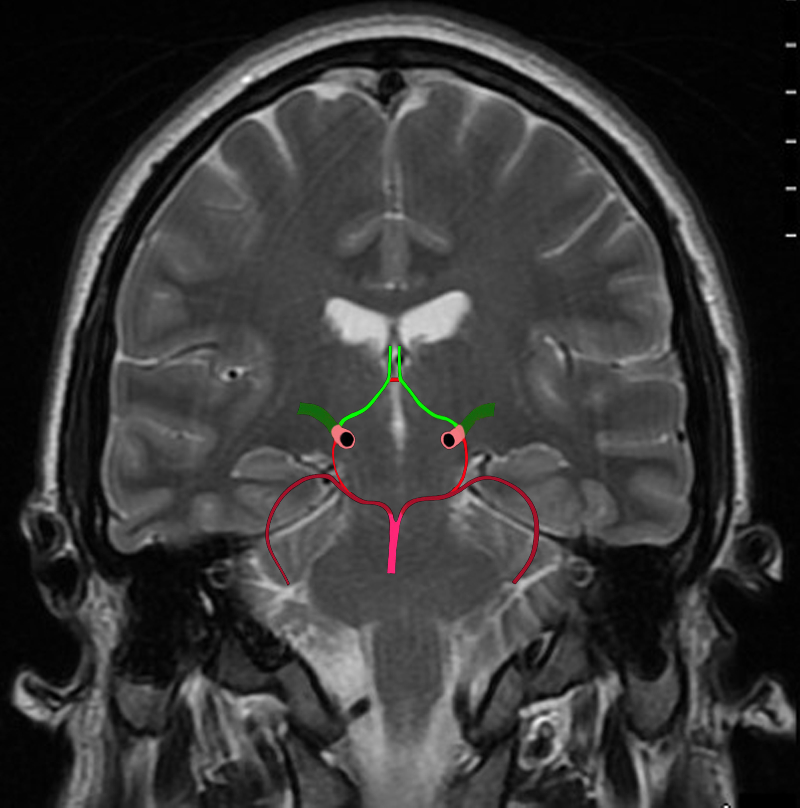
The Circle of Willis Overlaid on a Coronal View of the Brain |
|
The diagram shows the main branches of the blood supply to the brain including the circle of Willis overlaid on coronal MRI image to portray the approximate position of the vessels in the brain. The carotid system supplies the brain from the internal carotid we demonstrate its terminal bifurcation into middle cerebral (dark green) and anterior cerebral (bright green). The anterior communicating artery runs between the two anterior cerebrals (bright red) The basilar artery (pink) is formed by the two vertebral arteries and it travels as a single artery over the upper medulla and the pons. Its terminal branch is the posterior cerebral artery (maroon). Each of the carotid and vertebro-basilar systems contributes to the circle of Willis through communicating arteries. The vertebro-basilar system provides the posterior communicating arteries bilaterally from the posterior cerebral and the carotid system provides the anterior communicating arteries via the anterior cerebral arteries. Courtesy Ashley Davidoff MD Copyright 2010 All rights reserved 89721c06b.8sg04.8s |
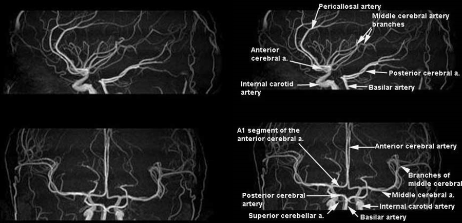
Map of the ACA and MCA |
|
The MRA in sagittal (upper images) and coronal projections (lower images) provide a map of the first and second order branches of the anterior and middle cerebral vessels. Image Courtesy of Philips Medical Systems 92484b.9s |
|
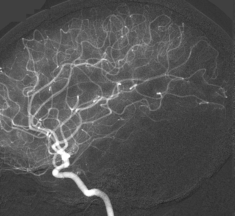
The Intracranial Circulation Derfinition of the Smaller Vessels Digital Angiogram of the Internal Carotid in the Lateral Projection |
|
The lateral digital angiogram of a selective internal carotid artery injection shows f the anterior and middle circulation supplied by the internal carotid artery. It is difficult to discern the origins of the anterior cerebral artery (ACA) and middle cerebral artery (MCA) in this projection. The study does however demonstrate the more distal portions of the ACA and MCA, and the smaller peripheral vessels. More specifically the loops of the distal middle cerebral arteries in the Sylvian fissure are well recognized. The intracranial portion of the internal carotid is well seen as well. Image Courtesy Ram Chavalli MD Copyright 2010 49420g01 |
|
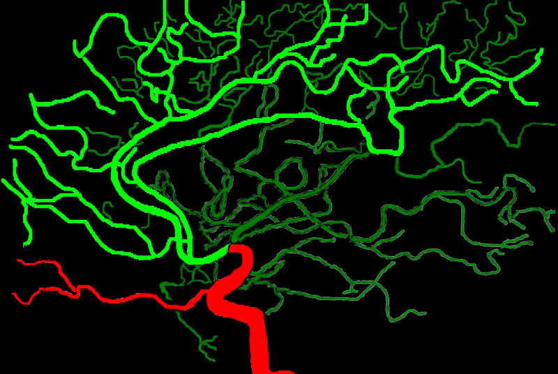
Anterior and Middle Cerebral Artery Distributions from a Lateral Perspective |
|
The anterior and middle cerebral circulation is demonstrated on the overlaid lateral angiogram of a selective internal carotid artery injection. The anterior cerebral circulation is overlaid in bright green and the middle cerebral circulation in dark green. The distal internal carotid artery and the ophthalmic artery (red) are shown (red) The ophthalmic artery is the smaller anterior horizontal vessel coursing to the orbit originates at the junction of the cavernous and supraclinoid segment. Image Courtesy of Philips Medical System Rendered by Ashley Davidoff MD Copyright 2010 All rights reserved 91390b01b04.8s |
|
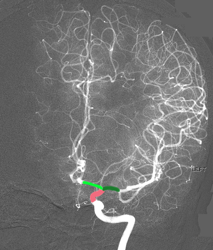
The Intracranial Circulation Derfinition of the Smaller Vessels Digital Angiogram of the Internal Carotid in the A-P Projection |
|
The intracerebral angiogram shows the normal appearance in the A-P projection of the anterior and middle cerebral territory. The supraclinoid portion of the terminal portion of the internal carotid artery divides into the anterior cerebral artery (AC – bright green) that courses medially and the middle cerebral artery (MCA- dark green) that branches laterally. The study does also demonstrates the more distal portions of the ACA and MCA, and the smaller peripheral vessels. More specifically the loops of the distal middle cerebral arteries in the Sylvian fissure are well recognized. The supraclinoid portion of the internal carotid is overlaid in salmon pink. Image Courtesy of Ram Chavalli MD 49422g01 |
Territory
Superficial branches: lateral frontal lobe, precentral gyrus, insula, postcentral gyrus, superior parietal gyrus, supramarginal gyrus, angular gyrus, temporal lobe.
Deep branches: lenticulostriate arteries, claustrum, external and extreme capsules; anterior ½ of the upper limb and the posterior limb of the internal capsule; putamen, globus pallidus, external caudate nucleus
|
Peripheral Branches of the Middle Cerebral Artery |
|
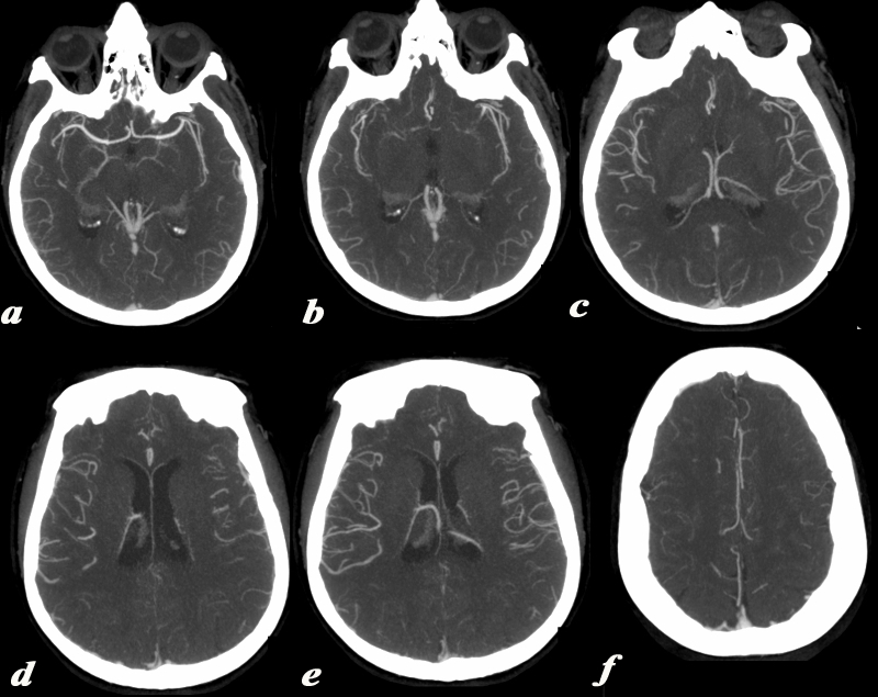
This series of axial images is from a CTA of the cerebral circulation and shows the middle cerebral artery at its origin (a) and its major branching pattern exemplified by the vessels branching in the Sylvian fissure to the frontal, temporal, and parietal lobes. The basal ganglia are fed by the middle cerebral vessels but they are relatively hypovascular compared to the cortex. The images proceed from inferior(a) to superior(f). Image Courtesy Ashley Davidoff MD Copyright 2010 v |
Middle Cerebral Arteries Highlighted |
|
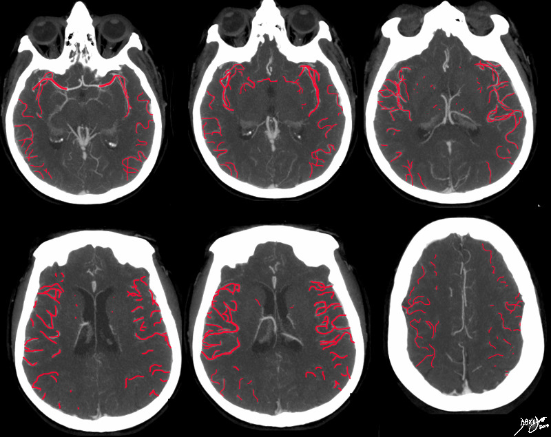
This series of axial images is from a CTA of the cerebral circulation and shows the middle cerebral artery at its origin (a) and its major branching pattern exemplified by the vessels branching in the Sylvian fissure to the frontal, temporal, and parietal lobes. The branches of the MCA have been overlaid in red. The basal ganglia are fed by the middle cerebral vessels but they are relatively hypovascular compared to the cortex. The images proceed from inferior(a) to superior(f). Image Courtesy Ashley Davidoff MD Copyright 2010 76500c03.8s |
Regions Perfused by the MCA |
|
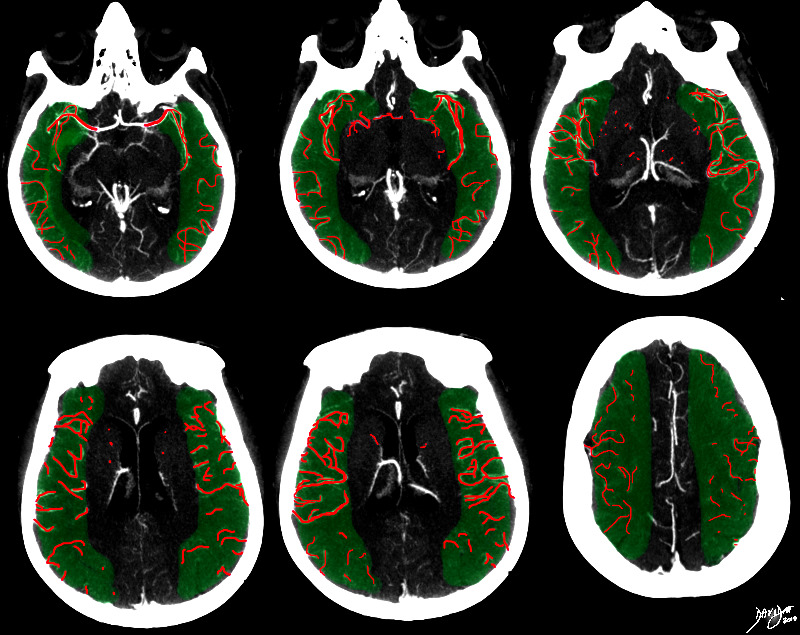
This series of axial images is from a CTA of the cerebral circulation and shows the middle cerebral artery at its origin (a) and its major branching pattern exemplified by the vessels branching in the Sylvian fissure to the frontal, temporal, and parietal lobes. The branches of the MCA have been overlaid in red, and the approximate territory perfused by these vessels overlaid in green. The basal ganglia are fed by the middle cerebral vessels but they are relatively hypovascular compared to the cortex. Image Courtesy Ashley Davidoff MD Copyright 2010 76500c04b.8s |
Perfusion territory of Anterior Middle and Posterior Cerebral Arteries |
|
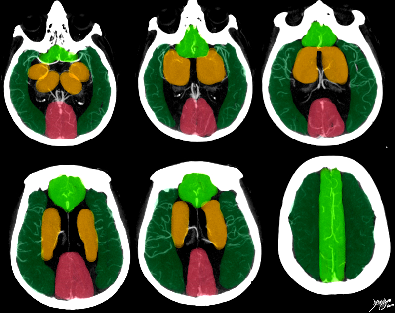
This series of axial images is from a CTA of the cerebral circulation and shows the middle cerebral artery at its origin and its major branching pattern exemplified by the vessels branching in the Sylvian fissure to the frontal, temporal, and parietal lobes. The branches of the MCA have been overlaid in red, and the approximate territory perfused by these vessels overlaid in dark green. The anterior cerebrl regions are overlaid in bright green and the regions of the posterior cerebral artery overlaid in salmon pink. The basal ganglia and thalamus are outline in orange and they receive most of their blood supply from the middle cerebral artery, but they are relatively hypovascular compared to the cortex. Image Courtesy Ashley Davidoff MD Copyright 2010 76500c02b.8s |
Disease
Anterior superficial MCA infarction (anterior ascending branches):
Hemiplegia (mostly face and arm); Broca’s aphasia (left lesion) ± dysarthria
Posterior superficial MCA infarction
Homonymous hemianopia; hemi-hypesthesia; Wernicke’s aphasia; apraxia; acalculia; visuospatial neglect (right infarct) ± hemiasomatognosia
Deep infarcts (perforating branches): hemiplegia, aphasia
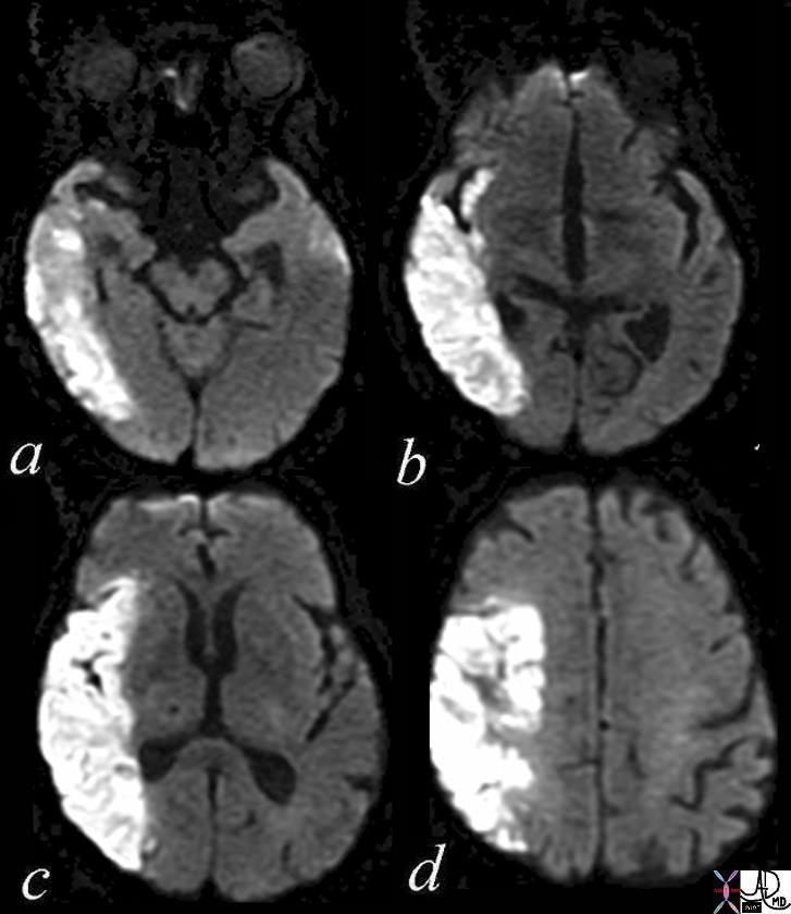
Right MCA Infarct – DWI |
|
The diffusion weighted MRI in axial projection shows a high intensity region in the right parietal and right temporal region revealing an acute infarction poattern in these regions reflecting right middle cerebral artery territory. Note that the basal ganglia have been spared, but the insula and operculum are involved. Courtesy Ashley Davidoff MD Copyright 2010 71275c01.800 71275c01c03 |
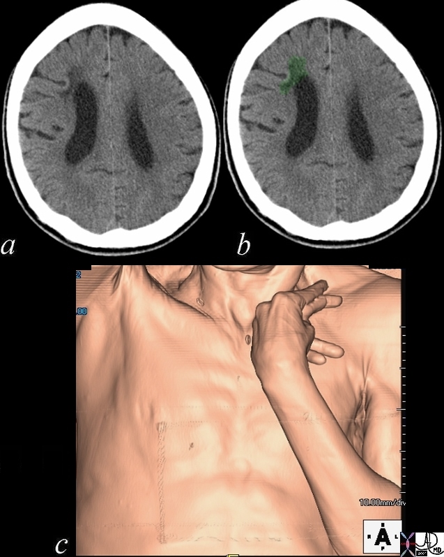
Chronic Frontoparietal Infarction |
|
This 60 year old male has the classical appearance of hypertonia of his upper limb and hand muscles as shown in this CT volume rendered image. The hand and elbow are flexed as a result of involvement on the contralateral motor cortex. The CT images show a hypodense region in the right frontoparietal region associated with encphalomalacia and ex vacuo changes in the right frontal horn consistent with a chronic stroke. Courtesy Ashley Davidoff MD 48641c01.800 |
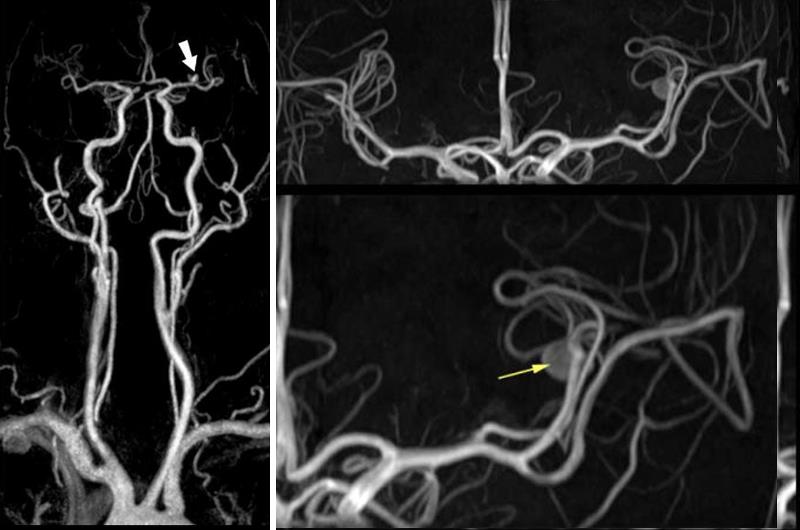
Left MCA Aneurysm – MRA |
|
The MRA performed on a 3T magnet shows an aneurysm of the left middle cerebral artery (MCA) demonstrated in the global frontal view (left image) by a large white arrow, and in the magnified view of the MCA (yellow arrow) Image Courtesy of Philips Medical Systems 92812.8 |
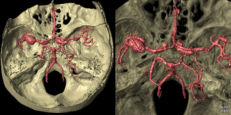
Right MCA Aneurysm and Ectasia of MCA’s Bilaterally CT – Volume Rendering |
|
The 3 D volume rendered image of the cerebral circulation looking into the middle and posterior cranial fossa with the circle of Willis in view shows a right sided middle cerebral artery aneurysm. Both middle cerebral arteries appear ectatic as well. Courtesy Philips Medical Systems 92579c |
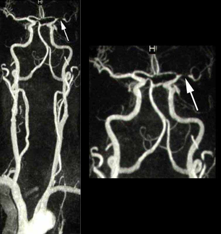
MRA of LMCA stenosis |
|
The MRA performed on a 3T magnet shows a stenosis (arrow) of the left middle cerebral artery (MCA) demonstrated in the global frontal view (left image) , and in the magnified view of the MCA Courtesy of Philips Medical Systems 92813c |
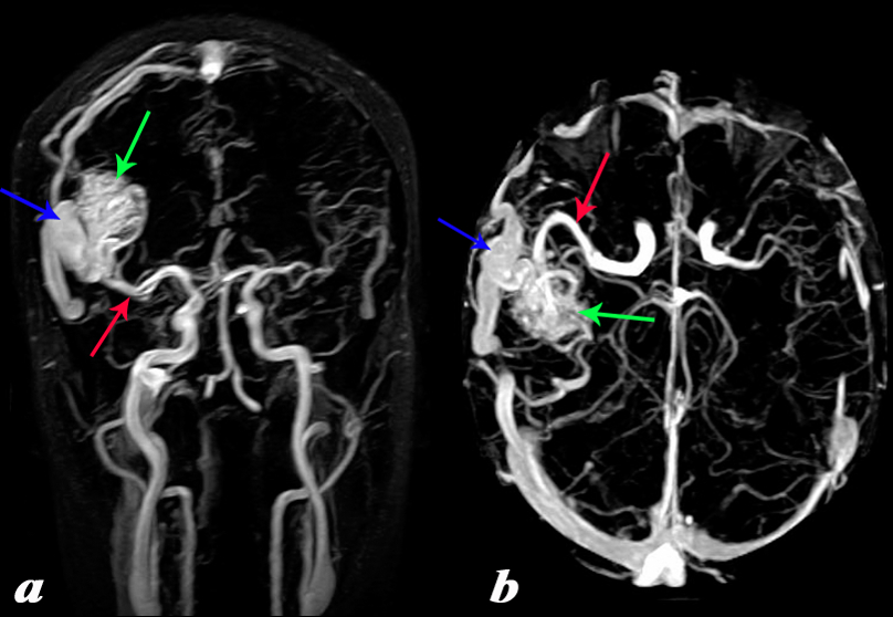
MCA AVM |
|
The MRA with 2d TOF in the coronal projection (a) and in the axial projection(b). It shows an arteriovenous malformation (AVM) in the right temperoparietal region fed by an enlarged middle cerebral artery (red arrow) and draining into a large Courtesy Philips Medical Systems 92465c01L.8 |
References
Slater D, Curtin S, Johns, J, Schmidt C. Middle Cerebral Artery Stroke e Medicine