
Cysticercosis
The Common Vein Copyright 2010
Definition Neurocysticercosis
Cysticercosis is a parasitic disease caused by ingestion of tapeworm eggs or its solium cysts by eating pork infested and undercooked. It is endemic in the tropics and subtropics. Neurocysticercosis refers to the infection involving the brain. It can result meningo-encephalitis, but mostly it is a syndrome of intracranial hypertension.
Infection is through ingestion of contaminated food or water contaminated. Contact with a carrier is an important risk factor. During the larval stage of tapeworm, the parasite can, through hematogeneous spread, implant and develop in various tissues of the body: skin, heart, eye, muscle, central nervous system.
The organism is present in tissues as cysts, which are fluid-filled, thin-walled and translucent. Depending on the host’s immune competence, the cysticercus calcifies more or less rapidly. The parasite usually lives 18 months to 2 years, but sometimes survives beyond 5 years.
The central nervous system is infected over 50% of cases. After the death of the parasite, the cyst starts to degenerate and then calcify. Parenchymal cysts, usually less than 1 cm, are located in the cerebral hemispheres. Intra ventricular cysts (10-20% cases) are found mostly in the fourth ventricle. Subarachnoid cysts may reach 10 cm or more.
Clinically, the telltale signs of the disease are variable. This may include seizures, from a mass of from cranial hypertension, deteriorating mental status, focal neurologic symptoms or stroke. Diagnosis can be made with serologies for antibodies against T. solium or circulating antigens of T. solium in serum and CSF.
CT and MRI are the examinations of choice. The images observed are not pathognomonic, but can distinguish the various locations and stages; typically, a lesion with central hypodensity of about 1cm is seen, with a regular peripheral rim, with surrounding edema.
Treatment is medical with praziquantel or albendazole. Surgery may be required in the presence of signs of intracranial hypertension, depending on the severity.
Cysticercosis by MRI |
|
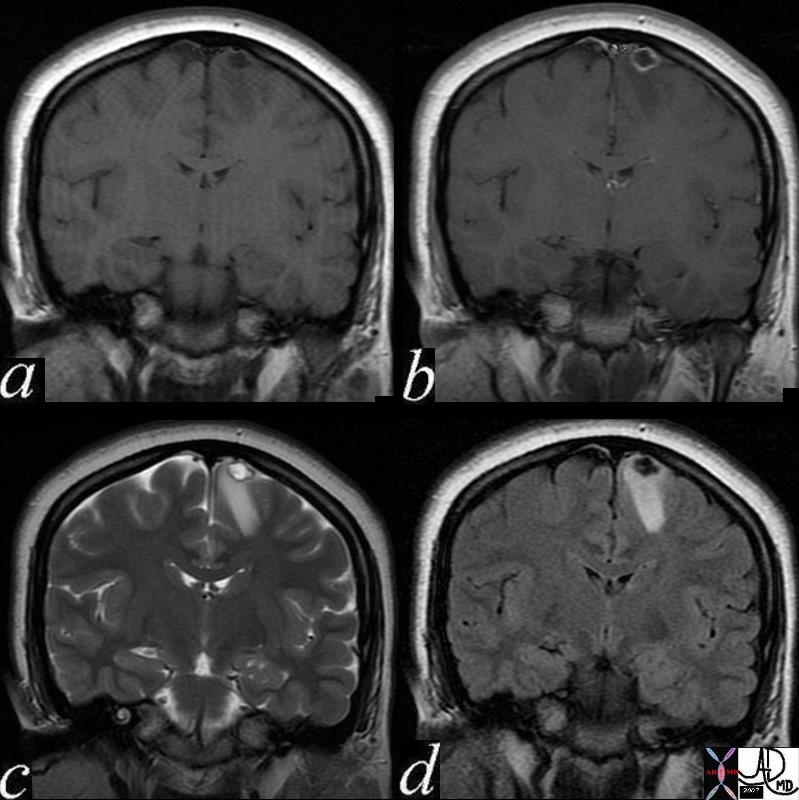
This 24 year old female presented with seizures An MRI with gadolinium enhancement suggested a diagnosis of cysticercosis. Image (a) is a pre gadolinium T1 weighted coronal image showing a hypointense focus in the paramedian part of the frontal lobe. Image (b) shows rim enhancement of the lesion. Image (c) is a coronal T2 weighted image that shows a hyperintense centre with a dark rim. A hypointense scolex can be seen as a tiny linear defect within the cavity. Surrounding edema is present in the white matter. Image(d) is a FLAIR coronal sequence and shows a dark matrix and edema in the surrounding white matter. The linear scolex inside the cavity is seen as relatively hyperintense. These finding are consistent with the diagnosis of cysticercosis. Courtesy Ashley Davidoff MD Copyright 2010 71575c02 |