
The Common Vein Copyright 2010
Definition
The parietooccipital sulcus separates the parietal lobe from the occipital lobe and is best appreciatesd in the sagittal plane
|
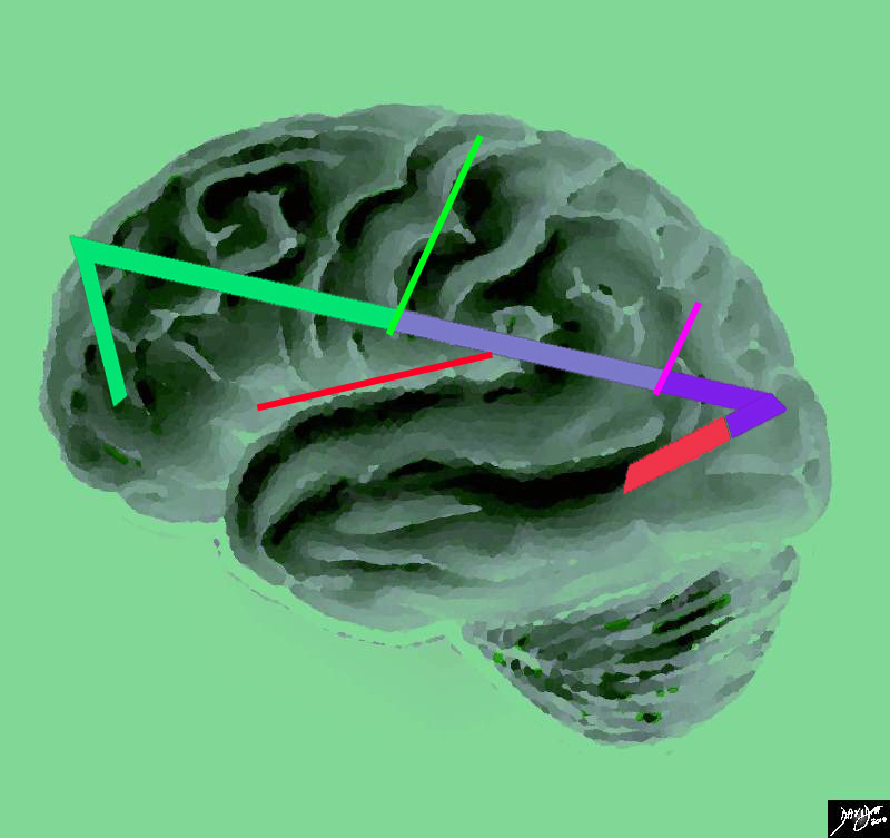
Central Sulcus in Bright Green |
|
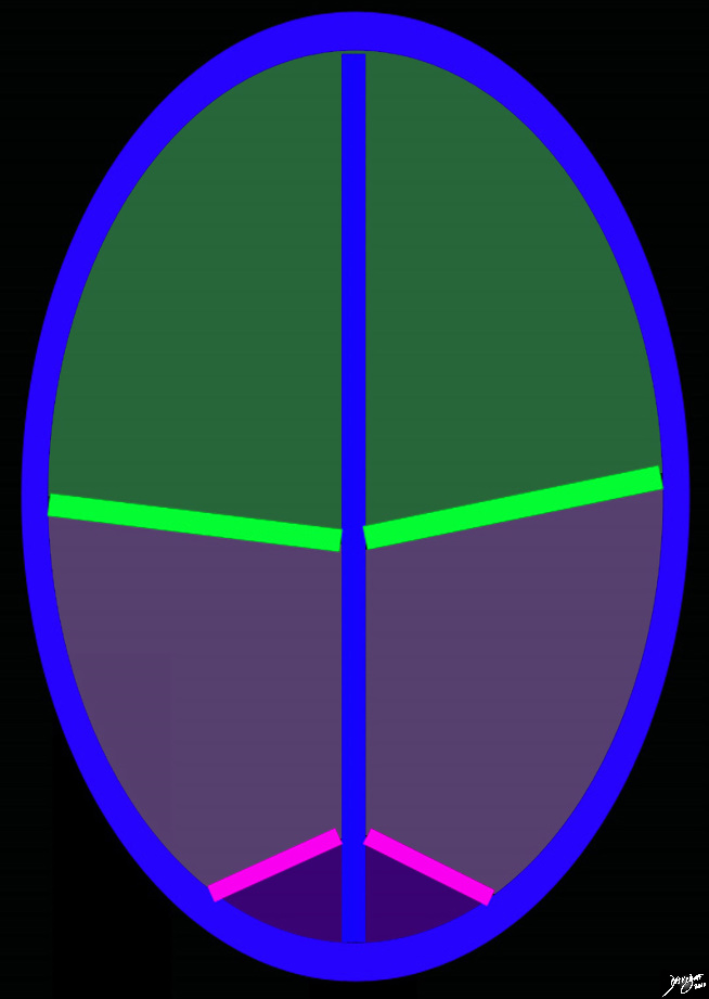
This artistic rendition of the brain reflects the vectors of the major parts of the brain revealing the major border forming fissures and sulci. The central sulcus (bright green line) divides the frontal lobe from the parietal lobe (light mauve). The region of the parieto-occipital fissure (pink line) divides the parietal lobe from the occipital lobe (purple). and the Sylvian fissure (thin red line)divides the temporal lobe from the frontal and parietal lobe Courtesy Ashley Davidoff copyright 2010 all rights reserved 83029e04.87s |
 Parieto-Occipital Fissure Parieto-Occipital Fissure |
|
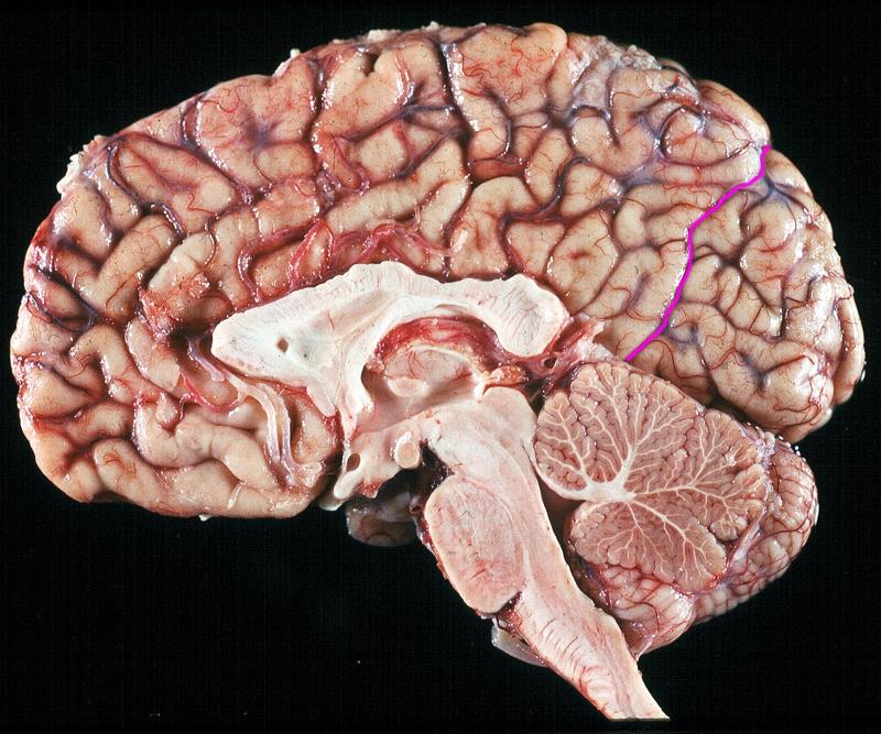
The sagittal view of the anatomical specimen of the brain shows the parieto-occipital fissure (pink) (aka sulcus), that separates the parietal lobe anteriorly and the occipital lobe posteriorly. Image Courtesy of Thomas W.Smith, MD; Department of Pathology, University of Massachusetts Medical School. 97805bd01 |
 Parieto-Occipital Fissure (pink) Parieto-Occipital Fissure (pink) |
|
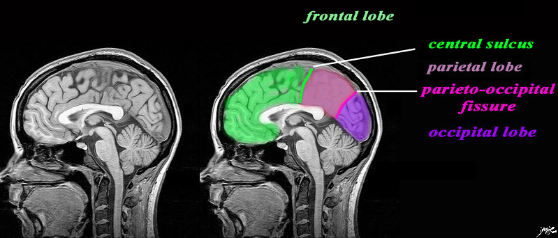
The sagittal image is deeper in the brain near the midline through the interhemispheric fissure, and is intended to demonstrate the the parieto-occipital fissure (pink) in order to define the border between the parietall and occipital lobe. The central sulcus (pink) has also been inferred in to demonstrate the border between the frontal lobe and parietal lobe. It is not usually Courtesy Philips MedicaL Systems rendered by Davidoff art 92141c01label.82s |
|
Occipital Lobe defined by the Parieto-Occipital Fissure |
|
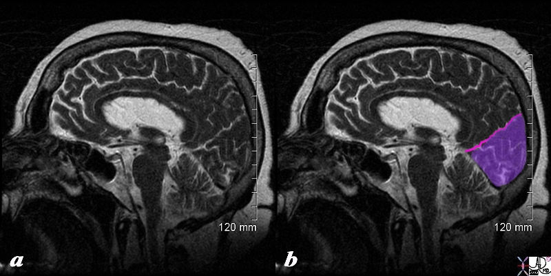
The sagittal view of the brain using a T2 weighted sequence shows the parieto-occipital fissure (pink) (aka sulcus), that separates the parietal lobe anteriorly and the occipital lobe (purple) posteriorly. Courtesy Ashley Davidoff copyright 2010 all rights reserved 71430cd01b.8s |
|
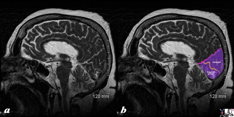
Occip[itoparietal Fissure Calcarine Fissure and Occipital Gyri |
|
The sagittal view of the brain using a T2 weighted sequence shows the parieto-occipital fissure (pink) (aka sulcus), that separates the parietal lobe anteriorly and the occipital lobe (purple) posteriorly. The calcarine fissure (orange) separates the cuneus above from the lingual gyrus inferiorly. Courtesy Ashley Davidoff copyright 2010 all rights reserved 71430cd01b02.8s |
|
Central Sulcus in the Axial Plane |
|
The diagram reflects the interhemispheric fissure as depicted in the axial plane dividing the brain into two halves. The bright green line represents the central sulcus which separates the the frontal lobes (green) from the parietal lobes (light purple. The parito-occipital fissure (pink) separates the parietal lobes from the occipital lobes. By convention anterior is on the top and posterior on the bottom, while the patients right is to our left and vice versa for the patients left Copyright 2010 Courtesy Ash;ley Davidoff MD 93914.85sb |
 Parieto-occipital Fissure in Axial Plane Parieto-occipital Fissure in Axial Plane |
|
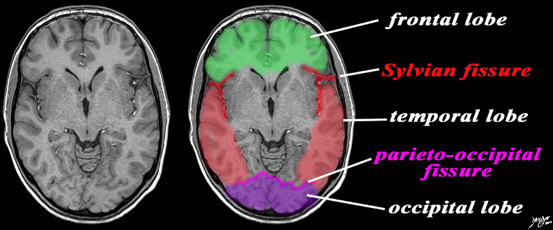
The axial image is intended to demonstrate ttwo important fissures; the Sylvian fissure (in red) the parieto-occipital fissure (pink) in order to demonstrate the border between the frontal and temporal lobe in the axial plane and the junction of the temporal and occipital lobe Image Courtesy Philips Medical systems rendered by Davidoff MD 92142c06b01 label.8s |