
The Common vein Copyright 2010
Introduction
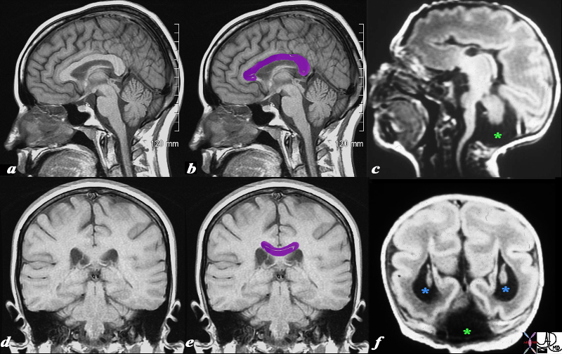
Normal (a,b,d,e) with Lissencephaly Associated with Dandy-Walker Syndrome and Agenesis of the Corpus Callosum |
|
This T1 weighted MRI is from a normal patient juxtaposed on a T1 weighted MRI from a neonate with agenesis of the corpus callosum, congenital lissencephaly and Dandy Walker syndrome. The normal corpus callosum (purple overlay ) is seen above the ventricles between the ventricles and the supracallosul gyrus and cingulate gyrus. . In the neonate (c,f) the there is no white matter in the expected position of the corpus callosum. These findings are consistent with a diagnosis of agenesis of the corpus callosum In addition, the posterior fossa does not show a normal cerebellum. Instead it is mostly filled with CSF (dark T1 green asterisk) likely due to a dilated 4th ventricle and atrophy of the vermis. Only a small amount of posterior fossa soft tissue is seen. In addition the posterior horns are dilated (blue asterisk). This finding is consistent with a diagnosis of Dandy-Walker syndrome associated with hydrocephalus. Lastly the patient has fewer gyri and sulci than normal. The gyri that are present are also more shallow than normal. These findings are consistent with lissencephaly. Courtesy James Donnelly MD Copyright 2010 All rights reserved 95466c01L03.8s |