
The Common Vein Copyright 2010
Definition
Dural sinus thrombosis is a circulatory abnormality of the dural sinuses characterized by thrombosis usually causedby a hypercoaguable state occurring in conditions such as protein C or protein S deficiency, use of oral contaraceptives, polycythemia vera nephtrotic syndrome, inflammatory bowel disease, and collagen vascular diseases such as SLE and Behcets disease. Local inflammatory disease such as meningitis, sinusitis, mastoiditis and surgery in the region are also associated causes.
The functional result may be focal ischemia due to venous ischemia or infarction but if sufficiently extensive global inhibition of venous drainage may result in elevation of intracranial pressure.
Clinically the patient may present with symptoms of elevated intracranial pressure such as headache, nause, vomitting visual disturbance or focal ischemic symptoms with motor or sensory disturbance.
The diagnosis is based on finding thrombosis of the sinuses on contrast enhance CT and or MRI.
Treatment is by anticiagualation unless there is significant associarted hemorrhage.
Structural Principles
|
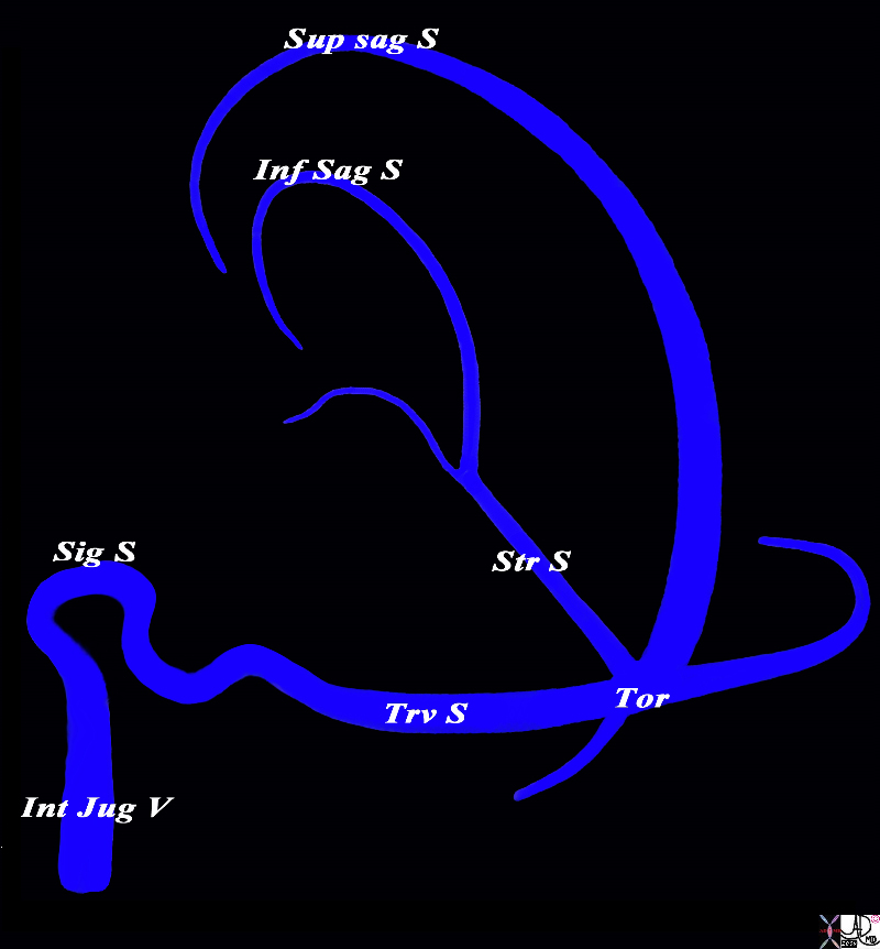
The Major Sinuses |
|
The diagram shows the basic plan of the dural sinuses and venous drainage of the brain in the parasagittal plane The superior sagittal sinus runs on the superior aspect of the falx and abuts the vertex of the skull. It usually empties into the right transverse sinus. The inferior sagittal sinus runs on the inferior aspect of the falx and empties into the straight sinus which in turn tends to empty into the left transverse sinus The transverse sinuses in turn empty into the sigmoid sinuses which exit the skull to enter the internal jugular veins. The confluence of the veins near the internal occipital protuberance is called the torcular heophili. Courtesy Ashley Davidoff MD Copyright 2010 All rights reserved 98057d01L018s |
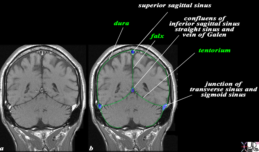
The Relationship of the Dural Sinuses and The Dura in the Coronal Plane |
|
The overlay outlines the dural sinuses (blue) and the dura (green) in this coronal T1 weighted normal MRI sequence. The peripheral dural sinuses include the superior sagittal sinus, the laterally placed junction of the transverse sinus and straight sinus, and the confluens of the inferior sagittal sinus which runs on the inferior surface of the falx and the straight sinus which lies on the posterior and superior surface of the tentorium overlying the cerebellum. The dura is outlined in green and it splits to enclose the dural sinuses and also forms the tentorium. The dura splits at the vertex to encompass the superior sagittal sinus and then extends down in the interhemispheric fissure to form the falx, splits again to encompass the inferior sagittal sinus anteriorly and straigt sinus posteriorly, and then spreads out to form the tentorium. The thin black T1 dark layer outward of the dura is the inner cortical bone, and is followed by the T1 bright fat containing marrow of the skull. Next is the thin black T1 dark outer cortical bone, followed by a thick layer of T1 bright subcutaneous fat, and then a thin layer of isointense (to soft tissue) skin layer. Image Courtesy Ashley Davidoff MD Copyright 2010 All rights reserved 98170c04.9Ls |