
The Common vein Copyright 2010
Definition
The tentorium is a 2mm horizontal layer of dura that extends from the occipital bone posteriorly extending and attaching to the petrous bones laterally being open anteriorly at the tentorial notch.
The tentorium cerebelli (aka tentorium, cerebellar tentorium) is an arched fold of duramater that is found between the anterior fossa and posterior fossa and forms the roof of the cerebellum, and the floor of the occipita;l lobes.
It looks like the roof of a tent and hence its name with the superior aspect being central and medial, and lowest points beig inferior and lateral.
Posteriorly it is attached to the occipital bone enclosing the transverse sinuses.
Anteriorly it has an oval opening called the tentorial notch which enables the midbain to pass through.
It is tough durable and about 2mm thick.
It divides the brain into supratentorial and infratentorial structures and diseases.
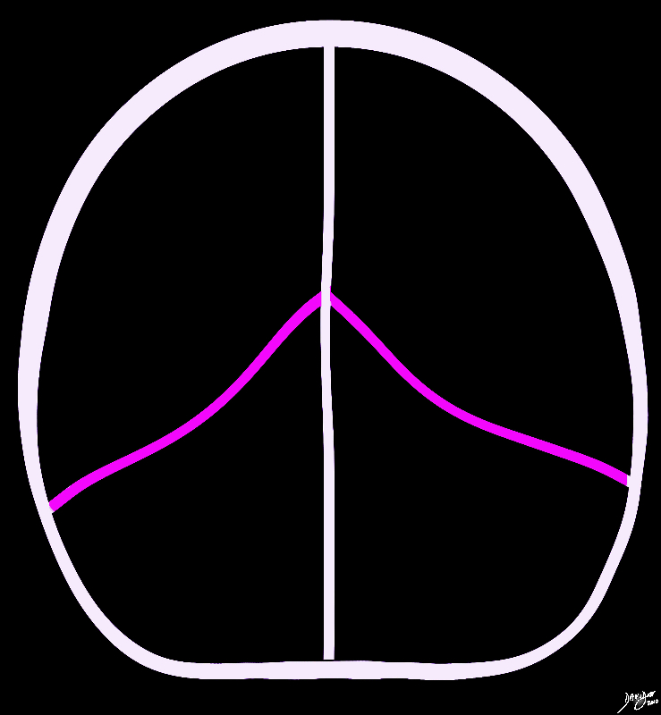 Coronal Diagram of the Tentorium Coronal Diagram of the Tentorium |
|
In this conceptual coronal drawing the distinction between the supratentorial and infratentorial space is made apparent by the bright pink tentorium that acts as a roof of the posterior fossa. The forebrain and the upper part of the midbrain lie above the tentorium, and the lower midbrain and hindbrain lie below. All the structures above the pink line are classified as supratentorial structures , and those below are infratentorial. Courtesy Ashley Davidoff MD copyright 2010 all rights reserved 93914b07g02.8s |
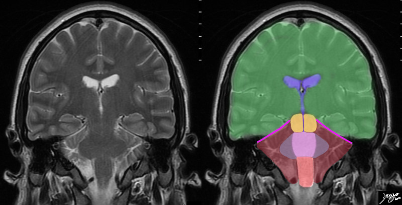 Coronal MRI tentorial relationship to Forebrain Midbrain and Hindbrain Coronal MRI tentorial relationship to Forebrain Midbrain and Hindbrain |
|
In this T2 weighted coronal MRI image the distinction between the supratentorial and infratentorial structures is made apparent by the bright pink tentorium that acts as a roof of the posterior fossa. The forebrain (green) midbrain (orange) and hindbrain (pink salmon and maroon) and the cerebellum (maroon), with other parts of the hindbrain filled in including the pons (light pink) middle cerebellar peduncles (mauve) and medulla (salmon) All the structures above the pink line are supratentorial, and those below are infratentorial. Part of the midbrain is supratentorial and part is infratentorial. The ventricular system is outlined in blue Courtesy Ashley Davidoff MD copyright 2010 all rights reserved 89721c06b.8sg01 |
|
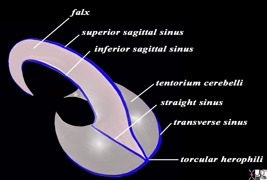
Dural Sinuses, Falx and Tentorium |
|
In this diagram the falx and the tentorium are exemplified together with their relationships to the dural sinuses. The image is drawn in a semisagittal plane showing the basic plan of the dural sinuses and relationship to the tentorium and falx. Note the sickle shaped falx posterior end having a greater craniocaudad dimension than the anterior end. The curved anterior aspect enables the falx to attach to the crista galli anteriorly as it proceeds under the frontal lobe. The tentorium cerebelli divides the brain into supratentorial and infratentorial structures. They are closed posteriorly and open anteriorly allowing the midbrain to pass through into the posterior fossa In the midline the straight sinus is enclosed and posteriorly the torcular herophili is present The superior sagittal sinus runs on the superior aspect of the falx and abuts the vertex of the skull. It usually empties into the right transverse sinus. The inferior sagittal sinus runs on the inferior aspect of the falx and empties into the straight sinus which in turn tends to Courtesy Ashley Davidoff MD Copyright 2010 All rights reserved 98057b09.9sL |
|
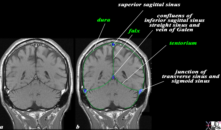
The Relationship of the Dural Sinuses and The Dura in the Coronal Plane |
|
The overlay outlines the dural sinuses (blue) and the dura (green) in this coronal T1 weighted normal MRI sequence. The peripheral dural sinuses include the superior sagittal sinus, the laterally placed junction of the transverse sinus and straight sinus, and the confluens of the inferior sagittal sinus which runs on the inferior surface of the falx and the straight sinus which lies on the posterior and superior surface of the tentorium overlying the cerebellum. The dura is outlined in green and it splits to enclose the dural sinuses and also forms the tentorium. The dura splits at the vertex to encompass the superior sagittal sinus and then extends down in the interhemispheric fissure to form the falx, splits again to encompass the inferior sagittal sinus anteriorly and straigt sinus posteriorly, and then spreads out to form the tentorium. The thin black T1 dark layer outward of the dura is the inner cortical bone, and is followed by the T1 bright fat containing marrow of the skull. Next is the thin black T1 dark outer cortical bone, followed by a thick layer of T1 bright subcutaneous fat, and then a thin layer of isointense (to soft tissue) skin layer. Image Courtesy Ashley Davidoff MD Copyright 2010 All rights reserved 98170c04.92Ls |
Applied Biology – Disease of the Tentorium
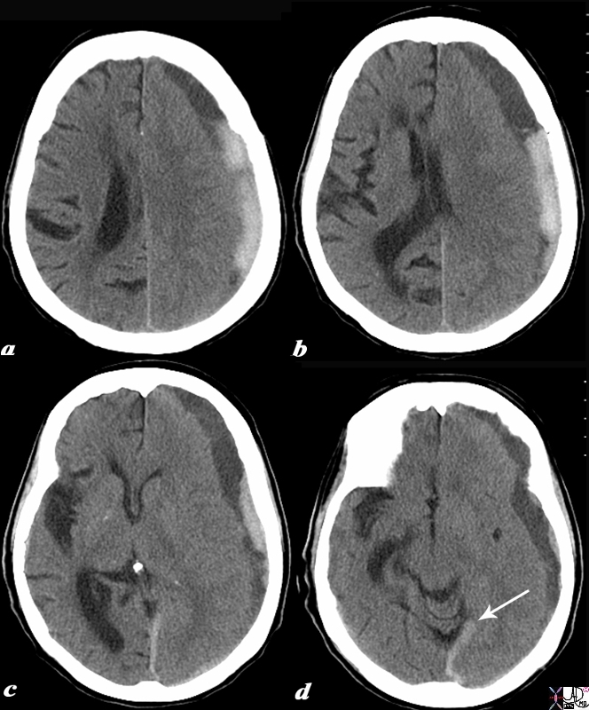 Acute on Chronic Subdural with Acute Component Extending Along the Free Margin of the Tentorium Acute on Chronic Subdural with Acute Component Extending Along the Free Margin of the Tentorium |
|
The CTscan without contrast is from a 88 year old male who has a history of falling who recently sustained head trauma and was complaining of a severe headache. The scan shows a collection in the left fronto-parietal collection with both high density and low density components. The left frontoparietal collection is subdural hematoma since it crosses suture lines, with the outer border conforming to the shape of the skull . Associated findings include flattening of the right sided frontal sulci and parietal sulci (a,b,c) as well as shift of the midline structures typified by the displacement of the septum pellucidum (b) and effacement of the left lateral ventricle(a,b,c). In image d, the acute hemorrhage has extended into the leaves of the posterior falx and inferior sagittal sinus (white arrow). The combination of low density and high density fluid in the subdural collection is consistent with an acute on chronic accumulation Courtesy Ashley Davidoff MD Copyright 2010 97737c.8s |