
The Common Vein Copyright 2010
Definition
A pituitary adenoma is a low grade neoplasm arising from the pituitary gland which may or may not be hormonally active. These adenomas are generally subcategorized by size, those under 1 cm are microadenomas and those over 1 cm are macroadenomas.
The etiology of pituitary adenomas is unknown but there may be some relationship to altered hormonal levels causing hyperplasia of the gland leading to growth and risk of tumor formation. There is a link between these masses and MEN type 1 and the Carney complex.
Pituitary adenomas are frequently incidental findings but may be present clinically by several mechanisms. Depending on which of the endocrine cell type the adenoma originated from, it can secrete any hormone produced in the anterior pituitary gland with the most common being prolactin. This can result in galactorrhea or amenorrhea. Macroadenomas may present clinically with endocrinologic abnormalities or with symptoms secondary to mass effect on adjacent structures such as the optic chiasm which can result in bitemporal hemianopsia. These lesions can also invade the cavernous sinus.
Diagnosis is generally made by a combination of imaging findings along with biochemical work up in the case of microadenomas. The diagnosis of nonfunctional macroadenomas may not always be straightforward as other masses may have a similar appearance, such as meningiomas.
MRI is generally more helpful than CT for identification of pituitary adenomas, particularly microadenomas. When seen, microadenomas are generally hypodense on CT, unless there is a component of hemorrhage. On MRI, they are generally T1 and T2 isointense to the rest of the gland and demonstrate relatively decreased enhancement. Associated findings are deviation of the pituitary stalk away from the side of the lesion and a convex superior margin of the involved gland. Macroadenomas have similar MRI signal characteristics but may demonstrate significant or heterogeneous enhancement. Increased T1 signal can be seen in hemorrhagic components of the mass.
Treatment for adenomas depends on many factors including size, hormonal alterations and invasiveness of the tumor. Treatment options include medical management, surgery or radiosurgery.
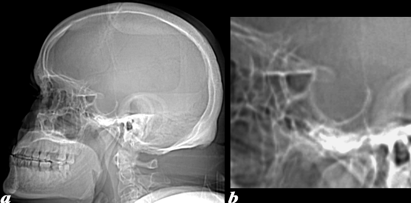
CT Scout of an Expanded Pituitary Fossa |
|
This 52 year old male presented with bitemporal visual field cuts. CT: On the lateral scout image from his CT, there is marked enlargement of the sella. Image a shows the enlarged pituitary fossa and sella turcica, and the fossa is magnified in image b. Image Courtesy Jimmy Wang MD 97677c.8 |
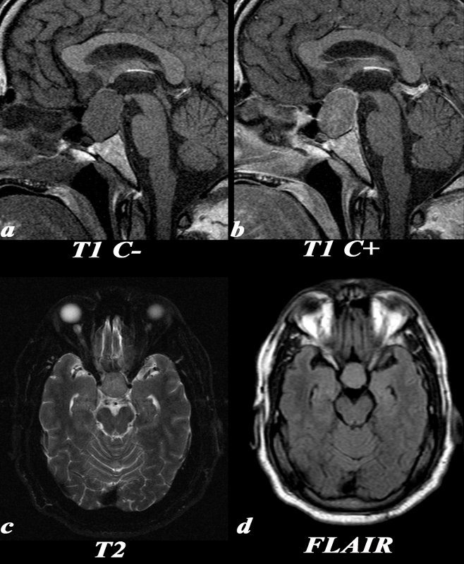
Homogeneous Tumor in the Pituitary Fossa |
|
This 52 year old male presented with bitemporal visual field cuts. T1 pre: This high resolution sagittal T1 weighted image through the sella demonstrates a relatively isointense rounded structure involving the sella and extending into the suprasellar area. The optic chiasm can be seen draped over the top and displaced superiorly by the mass. T1 post: Post contrast images demonstrate uniform enhancement of the mass. The thin rim of higher signal posteriorly and superiorly is most likely normal pituitary tissue stretched around the mass. Note that not all pituitary macroadenomas are this uniform in appearance. T2: Similar to the FLAIR image, there is an isointense to gray matter rounded structure in the suprasellar region. FLAIR: Notice the round, homogeneous lesion in the suprasellar region. It is isointense to gray matter. Image Courtesy Jimmy Wang MD 97678c.8 |
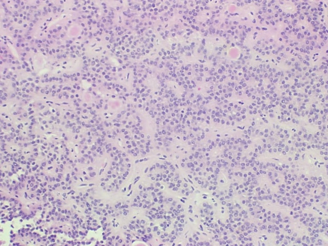
H&E Low Power
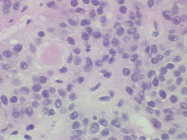
H&E High Power These low and high power polymicrographs demonstrate a sea of uniform cells arranged in gland like formation. A pathologic characteristic of pituitary adenomas is this uniform appearance, verses normal pituitary gland which is made up of numerous different types of cells producing various hormones.
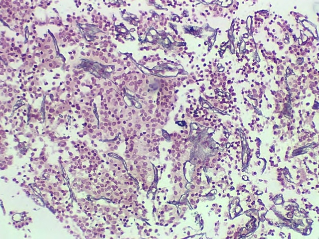
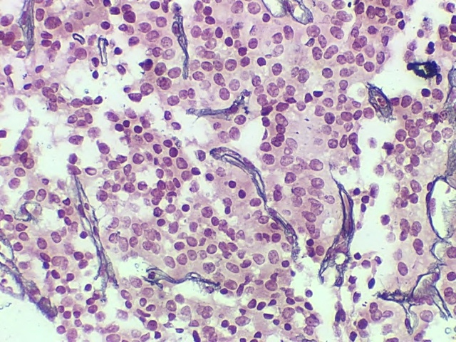
Reticulin Low Power
Reticulin High Power Pituitary Adenoma |
|
The low and high power polymicrographs are stained for reticulin . This stain is used to identify reticulin fibers which are identified within the architecture around normal pituitary tissue. In this case, the reticulin stain accentuated the blood vessels only, without staining around the cells seen in normal pituitary tissue. Image Courtesy of Cheryl Spencer, M.A. and Ivana Delalle, MD, PhD Department of Pathology Boston University School of Medicine 98516 /17/18/19 |
References
Requisites
AFIP notes