
Desmoplastic Infantile Ganglioglioma
The Common Vein Copyright 2010
Definition
Desmoplastic infantile ganglioglioma is a rare brain tumor which usually presents in infants under 1 year of age. It is usually large with an aggressive radiologic appearance but is generally a benign lesion. It is a heterogeneous tumor with both cystic and solid components which occupies more than one lobe with a predilection for the frontal and parietal lobes.
Pathologically, a desmoplastic infantile ganglioglioma is made up of desmoplastic stroma, with increased collagen-rich extracellular matrix from the adjacent neoplastic neural cells. Ganglion cells can also be identified within this mass.
Affected infants present with rapidly increasing head circumference and bulging fontanels.
Diagnosis is suspected based on imaging but confirmed on pathologic evaluation.
Imaging includes the use of both CT and MRI. On CT, these large lesions demonstrate both low attenuating cystic components and enhancing solid components. On MRI, the cystic components usually match CSF signal on T1 and T2 and are usually the deeper component whereas the solid component is peripherally located attached to the dura and demonstrates enhancement. Adjacent meningeal enhancement and thinning of the overlying skull has been seen.
Primary treatment of DIG is surgical resection, with complete resection being curative. Incomplete resection may be followed by adjuvant therapy.
|
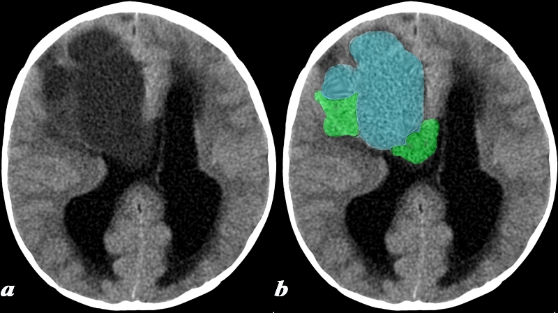
Cystic and Solid Components Causing Hydrocephalus |
|
This 17 month old boy presented with vomiting. CT: (a,b) A noncontrast head CT was obtained and demonstrates a predominantly cystic lesion (blue) with more solid components (green) at the periphery. Notice the enlargement of the lateral ventricles which is due to obstructive hydrocephalus caused by obstruction at the level of the third ventricle (not included on this image). These findings are consistent with a desmoplastic infantile ganglioglioma (DIG) Image Courtesy Elisa Flower MD and Asim Mian MD 97641c.8 |
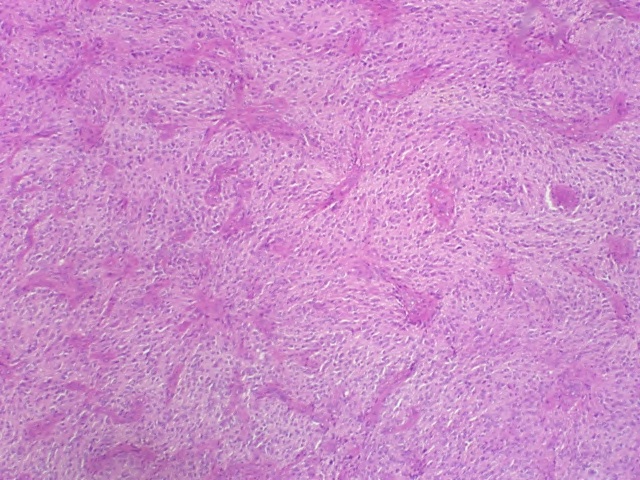
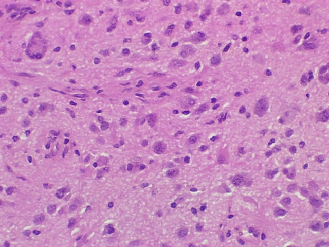
The low power polymicrograph demonstrates the overall vascular architecture of the tumor H&E
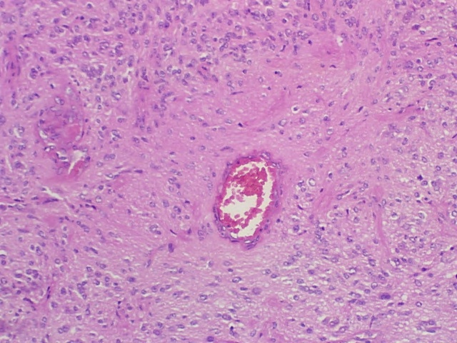
The medium power polymicrograph shows a vessel in the middle of the image without endothelial proliferation (HE4)(contrast this to this image of glioblastoma).
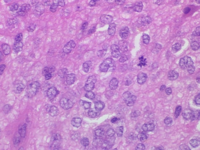
The high power polymicrograph demonstrates a mitotic figure, just to the right of center. Although mitoses are generally a predictor of more aggressive lesion, remember that patients with surgically resected desmoplastic infantile gangliogliomas have an excellent prognosis.
Another high power polymicrograph shows a multinucleated cell in the upper left region of the image which has the appearance of a giant cell but is actually a ganglion cell.
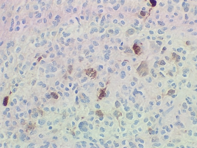
NeuN Marker Consistent with its classification as a mixed neuroglial tumor is positive staining for NeuN, a neuronal marker, and GFAP, a glial marker.
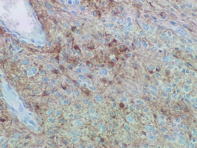
GFAP Glial Marker Consistent with its classification as a mixed neuroglial tumor is positive staining for GFAP, a glial marker.
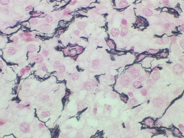
Staining for reticulin also demonstrates the surrounding connective tissue between the rests of tumor cells. DIG
|
|
Image Courtesy of Cheryl Spencer, M.A. and Ivana Delalle, MD, PhD Department of Pathology Boston University School of Medicine desmoplastic infantile ganglioglioma 98493/94/95/96/97/98/98b (S09-17273) |
References
Shin JH et al. Neuronal Tumors of the Central Nervous System: Radiologic Findings and Pathologic Correlation. Radiographic 2002; 22,1177-1189.