
The Common Vein Copyright 2010
Introduction
The cerebellum contains both gray and white matter. The folds of the cerebellum are narrow and deep and the cortex is distributed peripherally and the white matter centrally
The white matter consists of medullated nerve fibers whicgh pass out in radial fashion. They are of two types; those that originate in other parts of the nervous system, and those that orihginate in the Purkinje cells and connect with other parts of the nervous system.
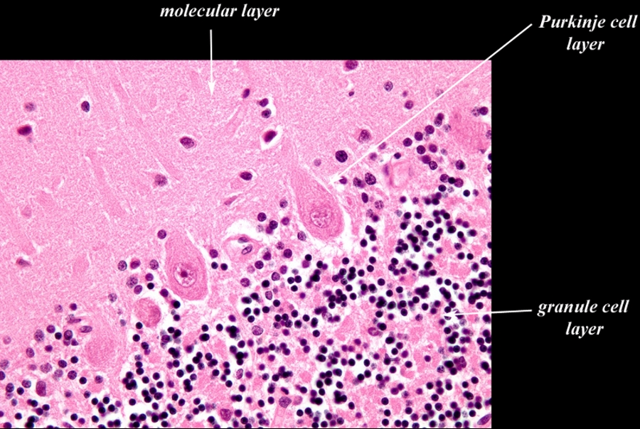
The cortex or gray matter consists of thrree layers;outer molecular layer, middle single celled layer of Purkinje cells and an internal granular layer.
The outer molecular layer contains larger and smaller multipolar cells.
 Cerebellar Gray Matter Cerebellar Gray Matter
3 Layers Inner Granule Cell LAyer Middle Purkinje and Outer Molecular Layer |
|
The image shows a histological specimen of the three layered cerebellar cortex typified by the large Purkinje cell in the single celled middle layer. The outer layer is called the molecular layer and contains larger and smaller multipolar cells. The inner layer is called the granule cell layer, granular layer or nuclear layer and is composed of closely packed cells dominated by the relatively nucleus and scant cytoplasm. Image Courtesy of Thomas W.Smith, MD; Department of Pathology, University of Massachusetts Medical School. 97375b01.9 |
The middle layer contains the cells of Purkinje which are large being some of the largest neurons in the brain, trumped only bythe Betz cells found in the primary motor cortex. The purkinje cells are integrative neurons of the cerebellar cortex. Each purkinje cell has innumerable branched dendrites which run perpendicular to the parallel fibers of the granule cells.
The internal granular or nuclear layer is composed of closely packed layer with cells dominated by the nucleus. The granule layer contains granule cells and golgi cells. Granule cells connect with the Purkinje cells.