Assistant
Introduction
CT MRI
Aging the Hemorrhage on MRI
Hyperacute – minutes to hours
T1 – hematoma (deoxyHb) dark => isointense
T2 – hematoma (deoxyHb) dark=> isointense
Acute – 0-2 days
deoxyhemoglobin is in intact RBCs with surrounding edema
T1- hematoma isointense, edema dark
T2 – hematoma dark, edema bright
Subacute – 2-14 days
deoxyhemoglobin changes to methemoglobin from outer to inner
T1 – outer core bright
T2 – outer core bright due to shortened T1, longer T2
Chronic – 14 days
hemosiderin laden macrophages at periphery
T1 – inner core bright, rim dark
T2 – inner core bright, rim has low dark
Chronic – months later
hemosiderin laden macrophages at periphery
T1- mostly iso-/dark, rim is dark
T2- markedly dark rim – “blooms” with greater T2-weighting
|
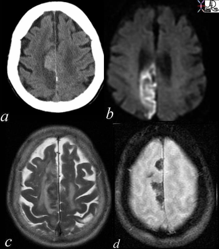
Acute Embolic Infarct High Parietal Region |
|
The axial images are from a patient with atrial fibrillation and neurological deficits. Image a is a CT scan which shows a high density lesion i the vertex of the right pariettal lobe suggesting hemorhagic change. Image b is a diffusion weighted MRI image at the level of the ventricles which shows a high intensity region in the parieto-occipital region suggesting acute infarction. Image c is a axial T2 weighted image showing edema in the white matter of the right parietal lobe. Image is aGRE image showing mixed heterogeneity with probable iron deposition suggesting subacute or chronic hemorhage. Findings are consistent with old and new multicentric infarcts of the brain likely from the heart caused by atrial fibrillation Courtesy Ashley Davidoff MD copyright 2010 71239c01 |
|
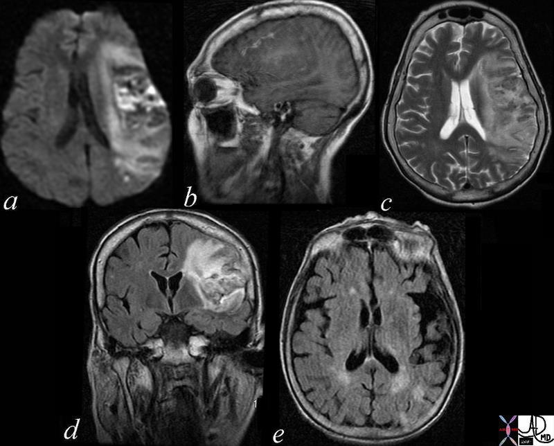
Subacute Hemorrhagic Infarct of the Parietal Lobe MRI |
|
The series of MRI images from a 70 year old male who by history suffered a stroke 1 month ago and has new onset symptoms. the series of images reveal complex changes of a subacute hemorrhagic infarct. Image a is from a DWI sequence and it shows a heterogenous increase in signal some of which represents T2 shine throughand sopme of which is bright raising the question on an acute on subacute entity. Image b is a sagittal T1 weighted image which shows areas of vague increase in density suggesting hemorrhage. Image c is a T2weighted sequence and shows some increase in water but the granular low intensity suggests hemosiderin deposit. Image d is FLAIR sequence showing increase brightness to the lesion in the left parietal lobe and image e is an axial FLAIR sequence The findings suggest extensive infarct in the left MCA territory which has mild mass effect on ventricles with petechial hemorrhage as seen on T2 and FLAIR and hyperintense T2 shine through on diffusion weighted images The punctate areas in left parietal lobe with restricted area of diffusion raises the question of a recent small infarct less bright regions on DWI suggests a subacute hemorrhagic infarct. Courtesy Ashley Davidoff MD copyright 2010 71000c03 |