Epidural Hematoma
The Common Vein Copyright 2010
Definition
When blood collects between the dura and the skull, most commonly due to arterial bleeding, an epidural hematoma is formed.
It is typically due to laceration of the middle meningeal artery, arising after trauma causing temporal bone fractures. Spontaneous hemorrhages and those resulting from acceleration-deceleration trauma are other possibilities. When a tear in middle meningeal artery happens, it may lead to a quick hemorrhage. Because blood would rapidly accumulate, patients can deteriorate within a few hours, the reason for which it is classically described as having a period of lucidity (loss of consciousness due to trauma, recovery and loss of consciousness due to mass effect – transtentorial herniation).
It can have a chronic presentation in about 10% of cases, which occurs when the vein, rather than the artery is the culprit for the hematoma formation.
Diagnosis is made by a CT scan, which shows blood (hyperdense), seen as a lenticular-shaped, convex mass. It does not cross the sutures. The hemorrhage displaces the falx and venous sinuses away from the inner table, being the only one that does so. MR allows the differentiation from acute and subacute bleeding, due to change in signal from methemoglobin (increasing signals in T1 and T2).
Treatment is with emergent surgical decompression and evacuation of the hematoma.
Structural Considerations
|
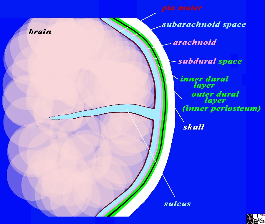
The Epidural Space Between the Outer Dural Layer (Inner Periosteum) and the Bone (Inner Table) |
|
The coronal drawing reveals the three layers of the brain. The inner layer (maroon) is the pia mater and it is in intimate contact with the brain and faithfully follows the sulci and gyri. The second layer is the arachnoid (pink) which is a slightly thicker membrane and follows the pia in a general fashion but does not extend into the sulci. It is intimately attached to the inner layer of the dura (bright green) The space between the pia and the arachnoid is the subarachnoid space, and it is in this space that the CSF is present and surrounds the brain. The next layer is the dura which is a double layer. The inner layer (bright green) is intimately attached to the arachnoid and the outer layer (also bright green) is attached to the bone and functions as the inner periosteum. There is a potential space between the arachnoid (pink) and the inner layer of the dura (green). This space is called the subdural space. (combination pink and green) The CSF (light blue) is seen in the subarachnoid space and in the lateral ventricles (gray blue) c Courtesy Ashley Davidoff MD Copyright 2010 All rights reserved 71422.800b02g06.91s |
|
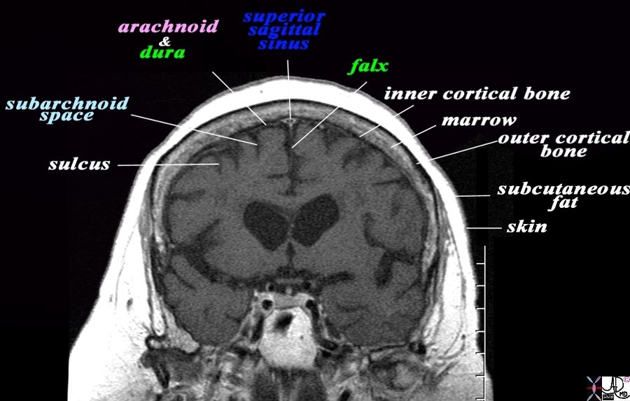
Dura and Arachnoid and Subarachnoid Space MRI T1 Weighted Coronal View |
|
The coronal T1 weighted MRI reveals demonstrates some of the anatomical features of the meninges and their relations The sulci are seen as T1 dark CSF containing fissures in the brain substance. The pia is too thin to see but would be adherent to the brain surface. The space that contains the CSF is called the subarachnoid space (light blue). The very fine T2 bright membrane seen on the surface of the brain is a combination of arachnoid (pink) adherent to the two layers of the dura (green). The outer layer of the dura acts as the inner periosteum and is adherent to the skull. The dura splits at the vertex to encompass the superior sagittal sinus and then extends down in the interhemispheric fissure to form the falx. The thin black T1 dark layer after the arachnoid and dura is the inner cortical bone, and is followed by the T1 bright fat containing marrow of the skull. Next is the thin black T1 dark outer cortical bone, followed by a thick layer of T1 bright subcutaneous fat, and then a thin layer of isointense (to soft tissue) skin layer. Image Courtesy Ashley Davidoff MD Copyright 2010 All rights reserved 71422.800bL01.9 |
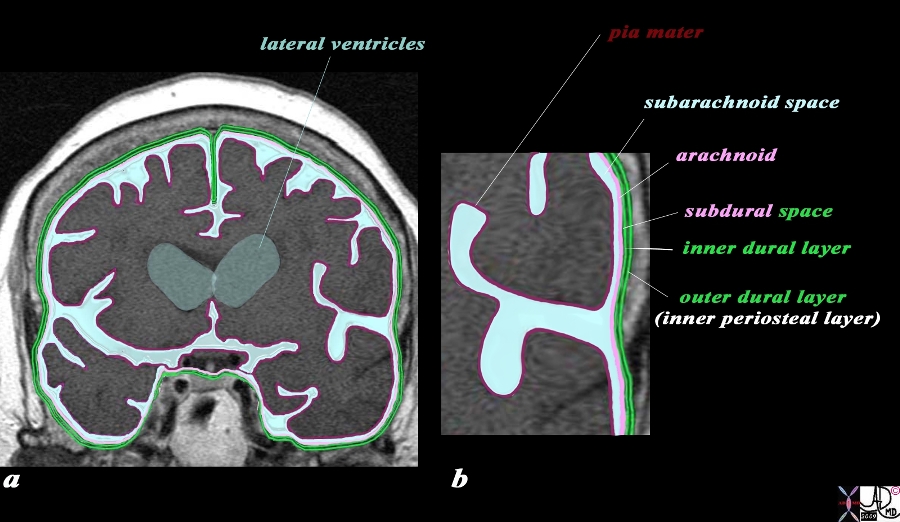
Dura and Arachnoid and Subarachnoid Space MRI T1 Weighted Coronal View |
|
The coronal image of the brain using T1 weighted MRI sequence reveals the three layers of the brain. The inner layer (maroon) is the pia mater and it is in intimate contact with the brain and faithfully follows the sulci and gyri. The second layer is the arachnoid (pink) which is a slightly thicker membrane and follows the pia in a general fashion but does not extend into the sulci. It is intimately attached to the inner layer of the dura (bright green) The space between the pia and the arachnoid is the subarachnoid space, and it is in this space that the CSF that surrounds the brain is housed. The next layer is the dura which is a double layer. The inner layer (bright green) is intimately attached to the arachnoid and the outer layer (also bright green) is attached to the bone and functions as the inner periosteum. There is a potential space between the two layers of the dura. The CSF (light blue) is seen in the subarachnoid space and in the lateral ventricles (gray blue) Image Courtesy Ashley Davidoff MD Copyright 2010 All rights reserved 71422.800b04c01.9s |
|
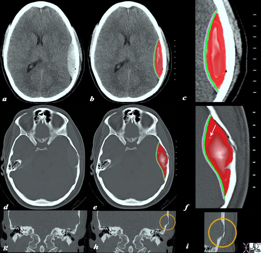
Epidural Hematoma Skull Fracture |
|
The CTscan without contrast is from a 19 year old male who sustained head trauma and who is complaining of a severe headache. The scan shows a high density lenticular shaped hematoma (a,b,c),. The right parietal collection is an extradural or epidural hematoma, with the outer border conforming to the shape of the skull and lined by the bone) and the inner border lined by two layers of dura (green c,f) and the arachnoid (pink c,f). There are bubbles of air scattered along the inner bone surface (black bubbles best seen on the magnified views (c, f) a tiny fragment of bone (white arrow in f) but seen on images d, and e as a white speck. The fracture of the temporal bone is ringed in orange on the coronal reconstructions of the axial views (h,i) Courtesy Ashley Davidoff MD Copyright 2010 98056c04.9s |