
Adrenoleukodystrophy
The Common Vein Copyright 2010
Definition
Adrenoleukodystrophy is an inherited metabolic disease which is characterized by progressive demyelination of the white matter along with adrenal insufficiency.
It is caused by a defective enzyme – lignoceryl-coA-ligase – which leads to accumulation of cholesterol esters (impairment in oxidation of very long fatty acids) in the white matter, the adrenal cortex and the testicles. This is a X-linked recessive transmitted disease.
Structurally, there is severe degeneration of the myelin, in the cerebrum, brainstem, optic nerves, and sometimes spinal cord. Macrophages containing degradation products of myelin can be identified (PAS positive). Additionally, the adrenal cortex and testicles are atrophic and replaced with lipids and fibrous tissue. In the CNS, it usually starts in the occipital lobe, advancing through the internal and external capsules and centrum semiovale, spreading then throughout the remaining white matter.
Clinically, individuals affected (mostly boys due to the transmission mode), has a early onset of symptoms, starting at ages 3-5, which are due to the loss of white matter and adrenal insufficiency. The latter is remarkable for the skin hyperpigmentation that first appears on patients, giving them a bronzed tonality, reason for which the disease is also called bronzed sclerosing encephalomyelitis. Neurologically, loss of vision and hearing start occurring, along with ataxia, change in personality and mental deterioration. A specific laboratory marker exists – an increase in hexacosanoic acids in plasma. Besides that, as predicted, sodium and chloride are low, potassium is high, and cosyntropin test fails to raise cortisol.
Radiologically, CT shows symmetric hypodense lesions in the occipitoparietaltemporal white matter, with enhancing rims surrounding them. Latter, cerebral atrophy is noted. MRI shows compatible changes, with hypointensity in the same areas described in T1, with corresponding increase in signal in T2WI.
Treatment is limited. Adrenal replacement therapy and a monounsaturated fatty acid rich diet with very low long fatty acids are options that can prolong life. Bone marrow transplantation is experimental, but has been shown to stabilize the disease.
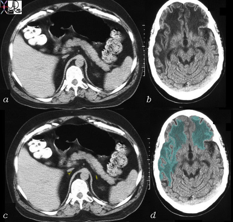
CTscan Adrenals and White Matter |
|
This CT is from a 26 year old male with known adrenoleukodystrophy. The findings on the CT of the brain (b,d) are characterised by significant changes in the white matter of both frontal lobes right parietal lobe and left parietal lobe to lesser extent. (green overlay) The adrenal glands (a,c) are barely visible but are overlaid in yellow in image (c). These findings are consistent with adrenoleukodystrophy. Courtesy Rebecca Schwartz MD Copyright 2010 All rights reserved 31464c04 |