
The Common Vein Copyright 2010
Definition
The lateral ventricles are two cavities that are arranged symmetrically on either side of the midline, extending longitudinally from the frontal to the occipital lobe. Each begins at the front lobe, head back to the posterior pole of the thalamus, surround it and then go forward again to the anterior temporal lobe.
At the posterior pole of the optic thalamus, where the ventricle changes direction, it originates a horizontal extension that merges into the occipital lobe. Each ventricle has therefore three parts: the anterior or frontal horn, the posterior or occipital horn, and an inferior horn (also called temporal or sphenoid). Moreover, the three poles converge in a single cavity, at the posterior end of the thalamus.
The lateral ventricles are separated from each other, and both communicate with the third ventricle through the foramen of Monro, located approximately in the middle of the anterior horn.
Shape
Curved shape, like a horseshoe – C-shaped – corresponding to the shape of each half of the brain.
Position
It passes through the four lobes of the brain.
The anterior horn is inferior to the corpus callosum, is separated from the opposite ventricle in the midline by the septum pellucidum and fornix, and is bounded laterally and inferiorly by the head of the caudate nucleus.
The posterior horn remains inferior to the corpus callosum and the optic tracts; it is superior to the medulla and splenium of the corpus callosum.
The inferior horn is inferior to the white matter of the hemispheres and tail of the caudate nucleus. It is superior to the hippocampus.
Character
Essentially a cavity whose walls are covered with ependyma, filled with CSF.
Part of the ventricular system in the brain, the lateral ventricles are filled with cerebrospinal fluid and connect to the central third ventricle. They are the largest of the ventricles. They are separated by the thin septum pellucidum. Disease in the lateral ventricles is manifested as hydrocephalus, and meningitis. There is also a relationship between schizophrenia and ventricle size. These diseases affect the entire ventricular system, not just the lateral ventricles. Pharmocological treatments are used as well as surgery to avoid the build up of excess CSF in the brain.
 Lateral Ventricles in Sagittal Lateral Ventricles in Sagittal |
|
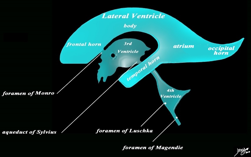
The diagram in the sagital projection reveals the horizontal portion called the lateral ventricle. It is a paired structure, and houses the frontal horn, body, occipital horn and atrium. The vertical portion consists of the paired foramina of Munro, the midline third ventricle, the narro aqueduct of Sylvius, the 4th ventricle the anteriorly placed paired foramina of Luschka, and theposteriorly positioned foramen Ashley Davidoff MD copyright 2010 all rights reserved 94459b10b02.82s |
 The Lateral Ventricles on a Variety of MRI Sequences The Lateral Ventricles on a Variety of MRI Sequences |
|
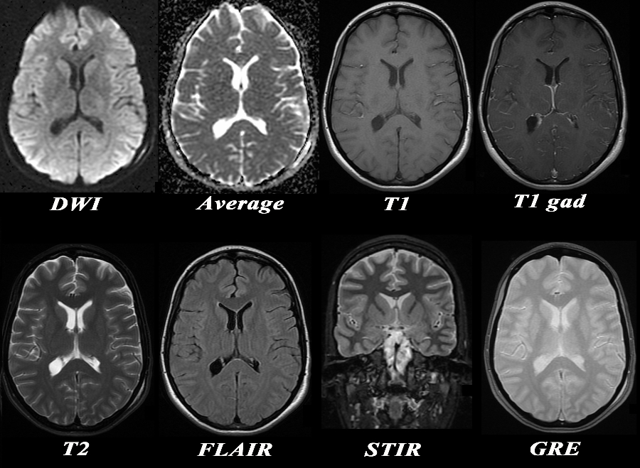
The MRI sequences used are displayed in the following graphic chart at the level of the lateral ventricles. The CSF is bright on the average diffusion images, the T2 weighted image, the STIR and the GRE sequence. The best differentiation of gray white matter is on the STIR sequences, while the basal ganglia are best seen on the STIR and GRE sequences Acute infarcts are best seen on the DWI images, enhancing tumors on the T1 with gadolinium and iron deposition of subacute or chronic hemorrhage best characterised on the GRE sequences. FLAIR sequences are sensitive to water in lesions and best characterize MS plaques Courtesy Ashley Davidoff MD Copyright 2010 95233c01.8 |
 Normal Lateral Ventricles of a Neonate by Ultrasound in Sagittal Plane Normal Lateral Ventricles of a Neonate by Ultrasound in Sagittal Plane |
|
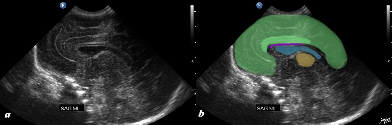
The ultrasound of a 7 day old baby boy reveals a normal sagittal section through the brain showing the CSF filled lateral ventricle (light blue) with most the brain substance showing characteristic homogeneous solid organ texture. The echogenic interfaces of the sulci with the gyri enable the outline of characteristic forms of the gyri so that for example the cingulate gyrus (darker green) is easily recognized by its classical location above the corpus callosum (purple). The corpus callosum is hypoechoic and overlaid in purple. The thalamus is isoechoic (orange) recognized by its position and shape rather than any unique ultrasound characteristics. The forebrain including the frontal parietal and temporal lobes are overlaid in green brain anatomy normal Lateral ventricle thalamus corpus callosum cingulate gyrus shape character US scan Neonate Ultrasound Courtesy Ashley Davidoff MD Copyright 2010 90986c02.8s |
 Lateral Ventricles Axial Lateral Ventricles Axial |
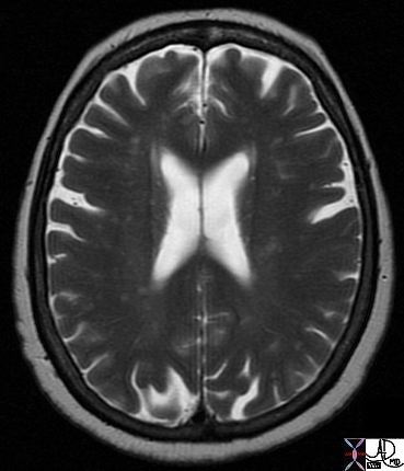
| Normal appearance of the lateral ventricles on a T2 weighted sequence. Since theey are fluid containing they are bright. Note the CSF in the sulci are also bright Courtesy Ashley Davidoff MD Copyright 2010 49047 |
Asymmetry of the Lateral ventricles – Normal Variant |
|
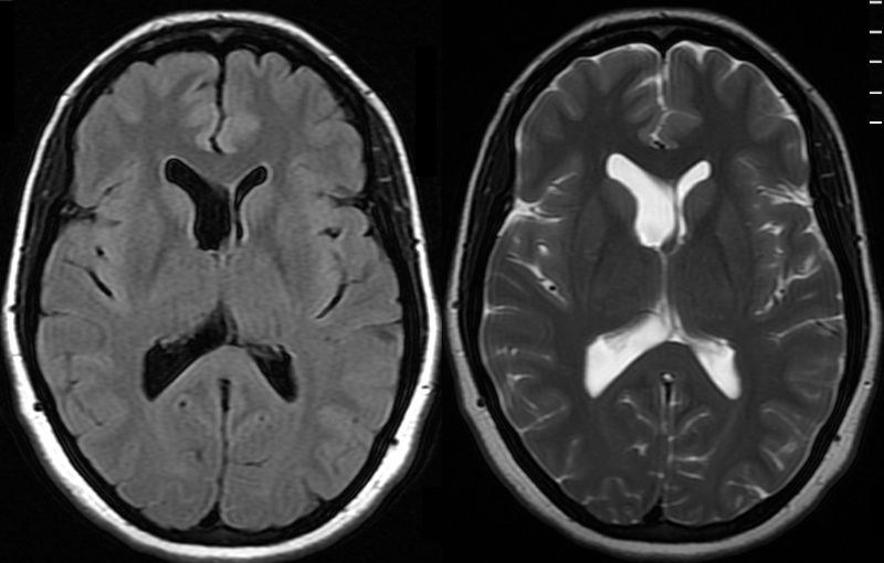
The MRI shows a FLAIR and a T2 weighted image revealing normal asymmetric development of the lateral ventricles in a 41 year old female. Courtesy Ashley Davidoff MD Copyright 2010 89044c |
Applied Biology Diseases
 70 year Brain in Two Different Patients “Normal” Involution (a) Normal Pressure Hydrocephalus (b) 70 year Brain in Two Different Patients “Normal” Involution (a) Normal Pressure Hydrocephalus (b) |
|
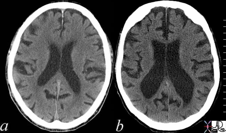
The 2 CT scans show 70 year old brains in two different patients. The patient on the left (a), reveals normal involution of the brain characterized by deepening of the sulci and prominence of the gyri together with a prominent ventricular system. Contrast this brain with the CT scan of the 22 year old above where the sulci and gyri are barely distinguished and the ventricles are slit like. The second patient (b) has a condition called normal pressure hydrocephalus (NPH). In addition to having “normal” involution, the patient also has dilated ventricles more than one would expect for the degree of involution. This is an abnormal finding. Courtesy Ashley Davidoff MD copyright 2010 72179c01 |
|
Acute Hemorrhage in the Left Fronto-parietal Region with Mass Effect on the Lateral Ventricles |
|
The brain has a characteristic appearance characterised by its creamy color and characteristic shape and fissured surface. This pathological specimen shows an acute hemorrhagic infarct in the left frontoparietal region with necrosis of the brain tissue and mass effect on the lateral ventricle and midline shift. The specimen also serves to reveal the normal right side with its creamy color and darker brownish gray matter that is accentuated by increasing the contrast on the right image (b). Other structures that are seen include the corpus callosum that lies above the ventricles (cc), the caudate nucleus (c) that lies inferior to the lateral ventricle, the line of white matter between the caudate and putamen called the internal capsule, the 3rd ventricle (3) medial to these structures , and the thalamus (th) inferior to them. The interhemispheric fissure (if) is seen superiorly and the Sylvian fissure (Sf) is seen laterally. The horizontal folds and darker color of the cerebellum are characteristics of that structure. Courtesy Ashley Davidoff MD Copyright 2010 10407c01 |

 Acute Cerebral Hemorrhage with Mass Effect and Rupture into the Ventricles Acute Cerebral Hemorrhage with Mass Effect and Rupture into the Ventricles |
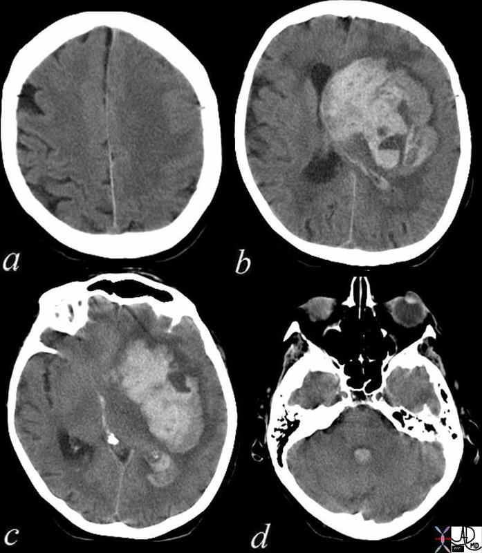
| This CT shows an acute hemorrhagic event originating in the frontoparietal region of the left cerebral hemisphere causing significant mass effect by compressing and displacing the ipsilateral lateral ventricle with significant midline shift.The hemorrhage has ruptured into the ipsilateral lateral ventricle and blood can be seen within the choroid plexus (b) in the posterior horn (c) as well as the 4th ventricle (d).
The ipsilateral edema has caused loss of the gray white matter interface in the left parietal lobe Courtesy Ashley Davidoff MD 72143c01 |
 Central Neurocytoma Central Neurocytoma
Classical Location in the Lateral Ventricle and associated with Hydrocephalus |
|
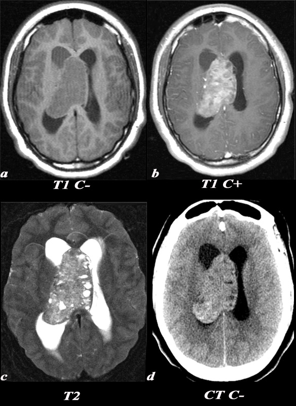
A 21 year old female was found to have papilledema on physical exam noted by her optometrist. MRI: T1 pre (T1 C- a) and post (T1 C+ b): Post contrast images demonstrate the heterogeneous enhancing nature of this mass. These images demonstrate centered in the right lateral ventricle with involvement of the septum pellucidum and partial extension into the left lateral ventricle. T2: T2 weighted images demonstrate internal areas of high signal consistent with cystic components. Also note the high T2 signal adjacent to the enlarged lateral ventricles which is a finding consistent with hydrocephalus, classically called transependymal flow of CSF. This unenhanced CT scan demonstrates also demonstrates the mass in the right lateral ventricle with similar features of heterogeneity and involvement of the septum pellucidum and partial extension into the left lateral ventricle. Notice the internal low density or cystic components and the higher density calcifications seen posteriorly. The lateral ventricles are enlarged consistent with resultant hydrocephalus. These findings are consistent with a central neurocytoma. Image Courtesy Elisa Flower MD and Asim Mian MD 97634c01.8s |