
The Common Vein Copyright 2010
Definition
The globus pallidus is part of the family of basal ganglia and lies between the putamen laterally and the head of the caudate nucleus medially.
Functionally it interacts with the substantia nigra and the dorsal striatum. (caudate nucleus and claustrum) It is divided by the medial medullary lamina. It functions as a pacemaker with characteristic firing rates and firing patterens.
It is targetted in treatments for Huntington’s disease.
|
The Globus Pallidus Nucleus (deep orange) Part of the Forebrain (Prosencephalon) Member of the Cerebrum (Telencephalon) Member of the Basal Ganglia Family
|
|
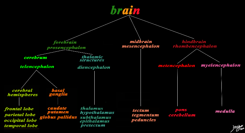
The basic and simplest classification of the brain into forebrain midbrain and hindbrain is shown in this diagram and advanced to a more complex tree using the embryological and evolutionary terminologies. The forebrain consists of the cerebrum also called the prosencephalon, which contains the more advanced form of the brain and the thalamic structures which contain more basic structures. The cerebrum (telencephalon) itself consists of two cerebral hemispheres and paired basal ganglial structures. Each cerebral hemisphere will have gray and white matter distributed in the frontal parietal temporal and occipital lobe, with the basal ganglia being part of the gray matter deep in the cerebral hemispheres. The most important thalamic structures arising from the diencephalons include the thalamus itself and the hypothalamus. The midbrain (mesencepaholon) consists of the tectum tegmentum and cerebral peduncles. The hindbrain has two major branch points based on the evolutionary development. The pons and cerebellum(part of the metencephalon) are grouped and the medulla (part of the myelencephalon is the second branch. Courtesy Ashley Davidoff MD Copyright 2010 All rights reserved 97686.8s |
 Globus Pallidus Forebrain Structure Globus Pallidus Forebrain Structure
member of the Structures that Abut the Ventricular System |
|
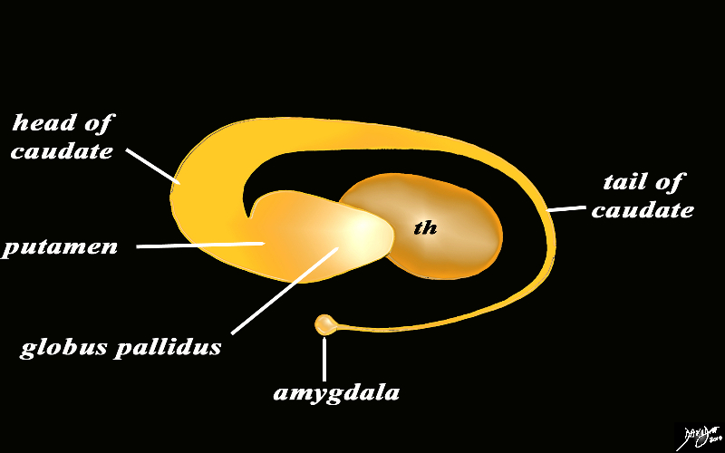
The orange c shaped ring lies lateral and to some extent inferior to the lateral ventricles typified by the light blue ring and consists of the thalamus and basal ganglia including the globus pallidus, putamen, head of the caudate nucleus, tail of the caudate nucleus and the amydala. This diagram infers the concept of recurring inverted C shaped structures that are layered both from deep to superficial but also to some extent medial to lateral. Courtesy Ashley Davidoff MD Copyright 2010 All rights reserved 93907d13b05b02.8s |
|
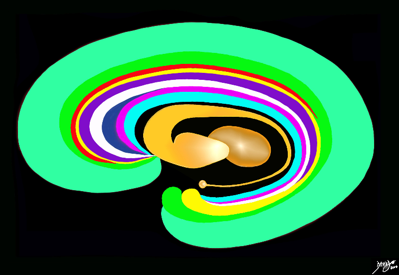
The Basal Ganglia of the Forebrain in Sagittal Projection Body and Tail of the Caudate |
|
The basal ganglia lie both in the forebrain and in the midbrain. In the forebrain they are distributed in paraventricular fashion almost in an inverted C fashion. In the conceptual diagrams we have colored the inverted “C” that abuts the ventricular system orange, and this includes the thalamus and the basal ganglia. The basal ganglia that lie in the forebrain include the globus pallidus, putamen, head of the caudate nucleus, tail of the caudate nucleus and the amygdala. The amygdala appears to be part of the limbic system and the basal ganglia. Courtesy Ashley Davidoff MD Copyright 2010 All rights reserved 93907d13b05dL3.8s |
|
The Globus Pallidus in the Context of the Other Basal Ganglia Axial Projection |
|
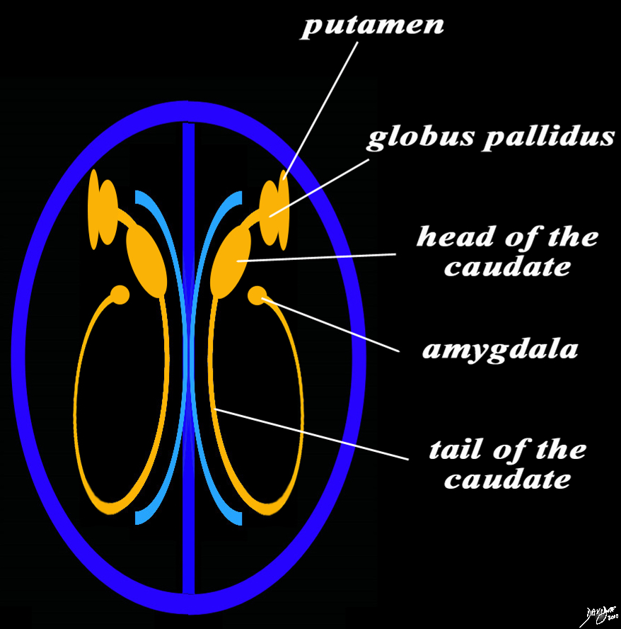
The basal ganglia in the forebrain in the axial projection are seen as a continuous inverted C as well. In this diagram their relationship to the ventricles distributed in paraventricular fashion is exemplified. In the conceptual diagrams we have colored the inverted “C” that abuts the ventricular system orange, and this includes the putamen, globus pallidus, head of the caudate nucleus, tail of the caudate nucleus and the amygdala. The amygdala appears to be part of the limbic system and the basal ganglia. Courtesy Ashley Davidoff MD Copyright 2010 All rights reserved 93914.3ka04b01e05.8s |

The Globus Pallidus Approximate Position on Axial CT Scan |
|
The basal ganglia in the forebrain in the axial projection on this CTscan are overlaid in dark orange while the thalamus is in lighter orange At a single axial level the major components of the basal ganglia of the forebrain are seen including the head and tail of the caudate nucleus, the putamen and the globus pallidus In this diagram their relationship to the ventricles distributed in paraventricular fashion (ventricles in blue) is also seen. Courtesy Ashley Davidoff MD Copyright 2010 All rights reserved 8568c12b08L |
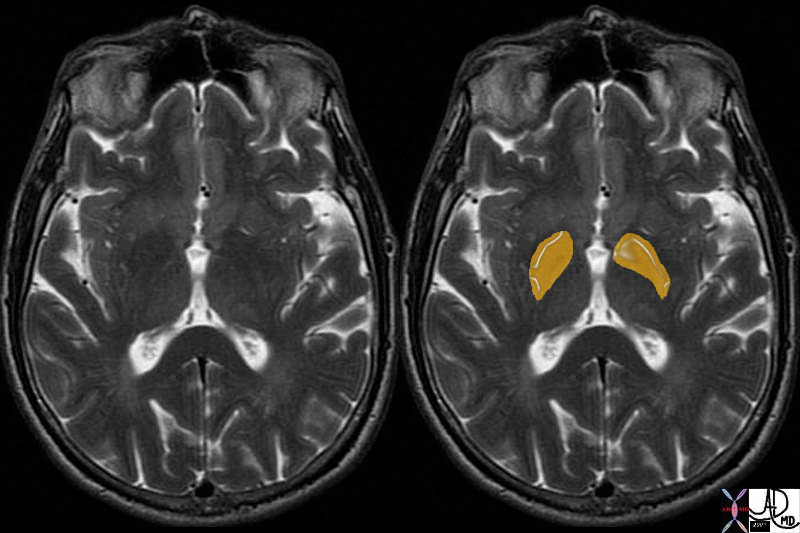 The Globus Pallidus The Globus Pallidus
T2 Weighted in the Axial Plane |
|
The globus pallidus is easily identified in the axial T2 weighted image lying medial and parallel to the putamen which is slightly more intense (contains more water) than the globus pallidus. The have the shape however both showing a type of comma appearance. Courtesy Ashley Davidoff MD Copyright 2010 38693c03 |
|
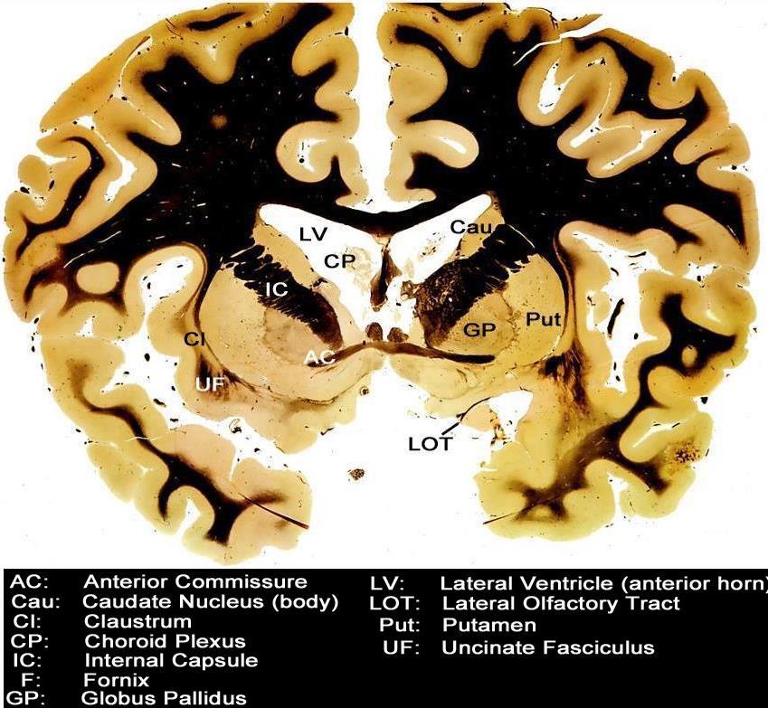
Coronal Section – Globus Pallidus (GP) |
|
This coronal section through the forebrain reveals the more superficial cortical structures that include the frontoparietal region and the temporal pole. Centrally the septum pellucidum surrounded by the choroid plexus (CP) is noted. The next paracentral structures include the left and right lateral ventricles (LV). The basal ganglia starting with the head of the caudate nucleus (Cau) and then the globus pallidus (GP and putamen (Put) follow, and these basal ganglia are separated by the internal capsule (IC). The claustrum (Cl) follow as the two lateral structures with the uncinate fasciculus (UF) lying inferiorly. The anterior commissure (AC) lie centrally and inferiorly and lateral tothe AC is the lateral olfactory tract (LOT). Courtesy Department of Anatomy and Neurobiology at Boston University School of Medicine Dr. Jennifer Luebke, and Dr. Douglas Rosene 97345.C6.8L01 |
|
Structural and Functional relationships of the Caudate Nucleus |
|
The coronal T1 weighted image reveals the structures that are functionally related to basal ganglial function. These include the caudate nucleus, globus pallidus, putamen, substantia nigra and subthalamus. The caudate nucleus and the putamen are the doorway to the basal ganglia and they receive input from both the sensory cortex and motor cortex. They distribute the signals to the globus pallidus substantia nigra and subthalamic nuclii. The latter (two subthalamic nuclii and substantia nigra) process the signal and send the result back to the globus pallidus which in turn sends the signal back to the thalamus. Courtesy Ashley Davidoff Md Copyright 2010 All rights reserved 38610c06c06d.81s |

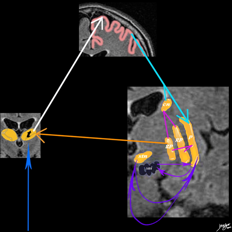 Integration of the Globus Pallidus and Other Basal Ganglia with the Sensory Cortex Integration of the Globus Pallidus and Other Basal Ganglia with the Sensory Cortex |
|
A simplified drawing of the connections between the caudate nucleus (orange c), the sensory cortex (salmon pink) and the basal ganglia is shown. After the stimulus has reached the sensory cortex for quantification and qualification it connects to the basal ganglia through the caudate nucleus and putamen. Each of these connects with the two parts of the globus pallidus (gp) which feed back to the thalamus. The caudate nucleus also feeds back and forth to the substantia nigra (sni) and the subthalamic nucleus (stn) putamen= p caudate nucleus = cn globus pallidus = gp substantia nigra = sni subthalamic nucleus = snu Davidoff art MRI T1 Courtesy Ashley Davidoff MD Copyright 2010 38610d09e02.8s |
References
José M. Hurtado, Charles M. Gray, Laszlo B. Tamas, and Karen A. Sigvardt Dynamics of tremor-related oscillations in the human globus pallidus: A single case study Proc Natl Acad Sci U S A. 1999 February 16; 96(4): 1674–1679.