Anterior Cerebral Artery
Copyright 2010
Origin
The anterior cerebral artery arises from the ventro-medial surface of the internal carotid artery. Its diameter (2-3 mm) is lower than that of the middle cerebral artery.
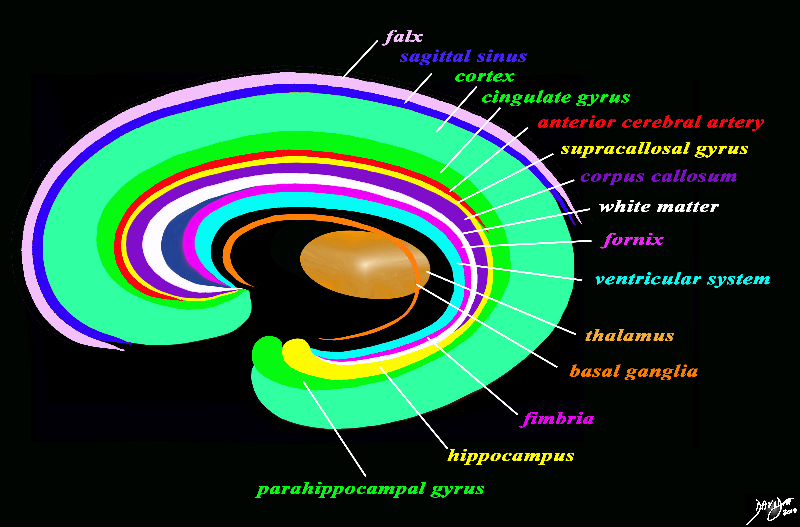
Part of the Rings |
|
The forebrain has most of its components aligned in a series of inverted c- shaped rings starting from the outer membranes that culminate in the falx (pink), then extending inward smaller inner rings with each intimately connected to the others. The thalamus (dull orange) appears diagrammatically as the centre of these rings as seen from the sagittal view The outer ring is the falx (pink) followed by the sagittal sinus (blue) cerebral cortex (light green), cingulate gyrus (bright green) superiorly which becomes the parahippocampal gyrus inferiorly. The red ring represents the distribution of the main portion of the anterior cerebral artery. Next is the yellow ring which is the supracollosal gyrus (indusium griseum) superiorly and the hippocampus inferiorly. This is followed by the corpus callosum (purple) which enables the white ring of white matter to connect between hemispheres. The next ring is the thin bright pink ring which represents the fornix superiorly and the fimbria inferiorly. The innermost ring (light blue) represents the lateral horns of the ventricular system. The basal ganglia run just lateral to the lateral ventricles. The navy blue arrow headed structure is the septum pellucidum. Courtesy Ashley Davidoff copyright 2010 all rights reserved 93907d13g03.8s |
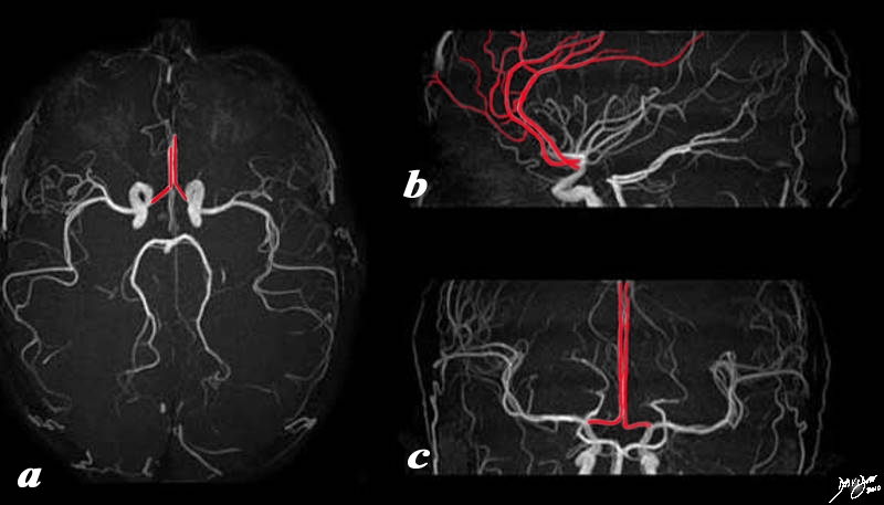
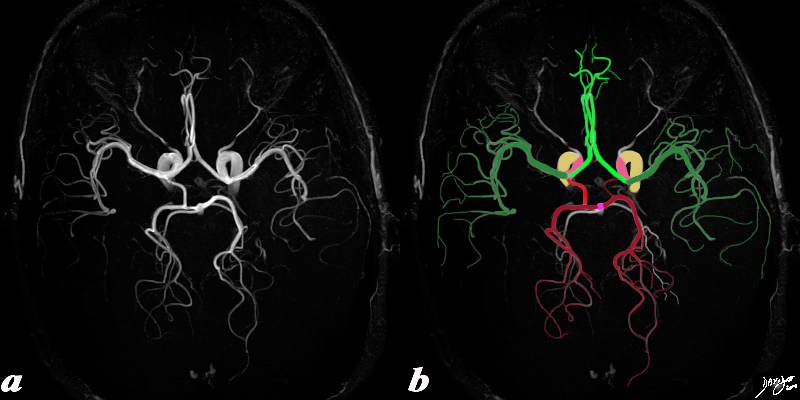
Overview of the Anterior Cerebral Artery in 3 Views – MRA |
|
There are two main branches off the internal carotid system as it enters the brain; anterior cerebral artery and the middle cerebral artery The MRA in 3 views shows the anterior cerebral artery overlaid in red. The best view to get a sense of its distribution is the lateral or sagittal view. On the axial view (a) the anterior cerebrals are seen arising as one of the terminal branches of the internal carotid artery. The vessels arise in close proximity and they run a course in parallel maintaining their close proximity. Their initial ascent is steep and then they turn posterior so only a small portion of the vessel is appreciated on a single axial view. On the sagittal view (b) the ascent anteriorly is noted to be almost vertical before it turns posteriorly conforming to the shape and form of the inverted C discussed in the organizational pattern of the brain.. In (c) the coronal view again shows the origin of the anterior cerebrals from the distal internal carotid and the close, vertical and parallel course that they run in their initial distribution. Image Courtesy Philips Medical Systems Artistic rendering Davidoff MD Copyright 2010 92479.83.8s |
Branches To and From the Circle of Willis |
|
This cerebral circulation is centered on the circle of Willis which has two basic systems that feed it; the carotid system (in this case salmon pink) and the vertebro-basilar system (brighter pink). They both feed into the circle of Willis (bright red) via communicating branches. The middle cerebral artery is the vessel that feeds into the COW by providing the anterior communicating artery and the posterior cerebral artery connects via the posterior communicating artery. Conceptually as depicted in this diagram the circle of Willis is the centre of the cerebral circulation and from it blood flows into the anterior (bright green), middle (darker green) and posterior cerebral (maroon) regions. Image Courtesy Ashley Davidoff MD Copyright 2010 97194b16b.8s |
|
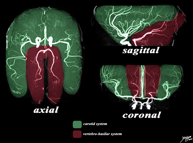
The Two Systems – Carotid and Vertebrobasilar |
|
There are two systems that supply the brain with blood. The MRA depicted in the axial, sagittal, and coronal views depict the general areas covered by the two systems. The carotid system gives rise to the anterior and middle cerebral arteries which cover the green area. The vertebro-basilar system, mostly through the posterior cerebral artery and cerebellar arteries, supply the maroon portion. Image Courtesy Philips Medical Systems Artistic rendering Davidoff MD Copyright 2010 92479c03.8s |
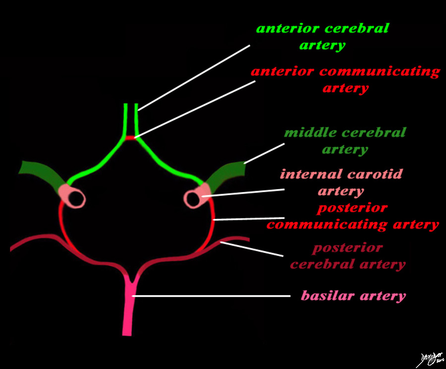
Structures of the Circle of Willis |
|
The diagram shows the main branches of the blood supply to the brain which includes the carotid and vertebro-basilar systems. These are the vessels that particpate in the formationofthe circle of Willis The carotid system supplies the brain from the internal carotid (salmon pink) – a branch of the common carotid which arises from the aorta. Its terminal bifuracation into the middle cerebral (dark green) and anterior green (bright green) are shown. The anterior communicating artery runs between the two anterior cerebrals (bright red) The basilar artery (pink) is formed by the two vertebral arteries and travel as a single artery over the upper medulla and the entire pons. Its terminal branch is the posterior cerebral artery (maroon). Each of the vessels contributes to the circle of Willis through communicating arteries. The vertebro-basilar system provides the posterior communicating arteries bilaterally and the carotid system provides the anterior communicating arteries via the anterior cerebral artery. code brain Courtesy Ashley Davidoff MD Copyright 2010 All rights reserved 97194b13g04L01.91s |
|
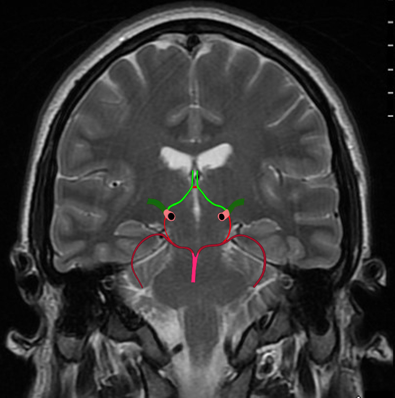
The Circle of Willis Overlaid on a Coronal View of the Brain |
|
The diagram shows the main branches of the blood supply to the brain including the circle of Willis overlaid on coronal MRI image to portray the approximate position of the vessels in the brain. The carotid system supplies the brain from the internal carotid we demonstrate its terminal bifurcation into middle cerebral (dark green) and anterior cerebral (bright green). The anterior communicating artery runs between the two anterior cerebrals (bright red) The basilar artery (pink) is formed by the two vertebral arteries and it travels as a single artery over the upper medulla and the pons. Its terminal branch is the posterior cerebral artery (maroon). Each of the carotid and vertebro-basilar systems contributes to the circle of Willis through communicating arteries. The vertebro-basilar system provides the posterior communicating arteries bilaterally from the posterior cerebral and the carotid system provides the anterior communicating arteries via the anterior cerebral arteries. Courtesy Ashley Davidoff MD Copyright 2010 All rights reserved 89721c06b.8sg04.8s |
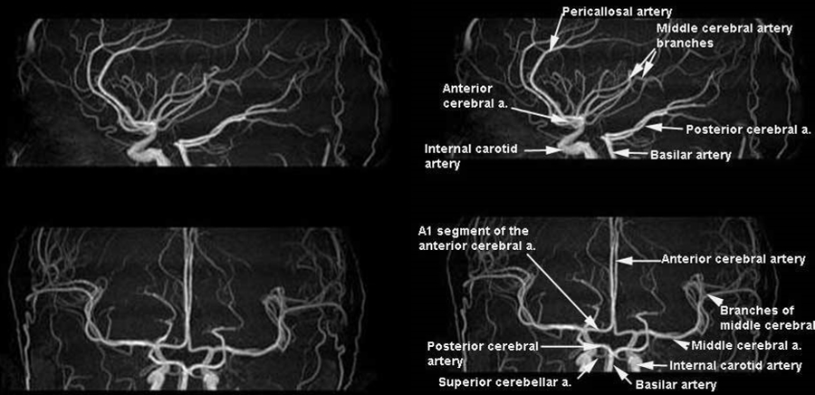
Map of the ACA and MCA |
|
The MRA in sagittal (upper images) and coronal projections (lower images) provide a map of the first and second order branches of the anterior and middle cerebral arteries. Image Courtesy of Philips Medical Systems 92484b.9s |
|
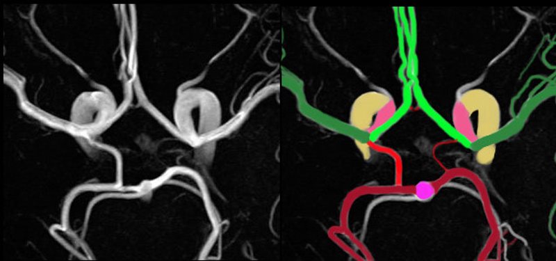
End of the Line for the Internal Carotid |
|
The axial MRA using 3T magnet shows a magnified view of the circle of Willis overlaid in color to reflect the component vessels. The supraclinoid portion of the internal carotid (salmon pink) divides into the middle cerebral (MCA dark green) and anterior cerebral (ACA bright green). There is a tiny anterior communicating artery (bright red) that runs between the two anterior cerebrals. The basilar artery (bright pink) gives rise to the posterior cerebral (PCA maroon) and superior cerebellar arteries (white) carotid system. The posterior cerebral artery gives rise to the posterior communicating artery which plugs into the region of the origins of the MCA and ACA. Note the left posterior communicating artery is relatively small which is a normal variant. Each of the carotid and vertebro-basilar systems contributes to the circle of Willis through communicating arteries. The vertebro-basilar system provides the posterior communicating arteries bilaterally from the posterior cerebral and the carotid system provides the anterior communicating arteries via the anterior cerebral arteries. Image Courtesy Philips Medical Systems Image Rendered Ashley Davidoff MD Copyright 2010 All rights reserved 92550c01.8s |
|
MRA of the Circle of Willis |
|
The axial MRA using 3T magnet shows a magnified view of the circle of Willis overlaid in color to reflect the component vessels. The supraclinoid portion of the internal carotid (salmon pink) divides into the middle cerebral (MCA dark green) and anterior cerebral (ACA bright green). There is a tiny anterior communicating artery (bright red) that runs between the two anterior cerebrals. The basilar artery (bright pink) gives rise to the posterior cerebral (PCA maroon) and superior cerebellar arteries (white) carotid system. The posterior cerebral artery gives rise to the posterior communicating artery which plugs into the region of the origins of the MCA and ACA. Note the left posterior communicating artery is relatively small which is a normal variant. Each of the carotid and vertebro-basilar systems contributes to the circle of Willis through communicating arteries. The vertebro-basilar system provides the posterior communicating arteries bilaterally from the posterior cerebral and the carotid system provides the anterior communicating arteries via the anterior cerebral arteries. Image Courtesy Philips Medical Systems Image Rendered Ashley Davidoff MD Copyright 2010 All rights reserved 92550c02.8 |
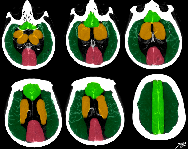
Perfusion territory of Anterior Middle and Poserior Cerebral Arteries |
|
This series of axial images is from a CTA of the cerebral circulation and shows the middle cerebral artery at its origin and its major branching pattern exemplified by the vessels branching in the Sylvian fissure to the frontal, temporal, and parietal lobes. The branches of the MCA have been overlaid in red, and the approximate territory perfused by these vessels overlaid in dark green. The anterior cerebrl regions are overlaid in bright green and the regions of the posterior cerebral artery overlaid in salmon pink. The basal ganglia and thalamus are outline in orange and they receive most of their blood supply from the middle cerebral artery, but they are relatively hypovascular compared to the cortex. Courtesy Ashley Davidoff MD Copyright 2010 76500c02b.8s |
|
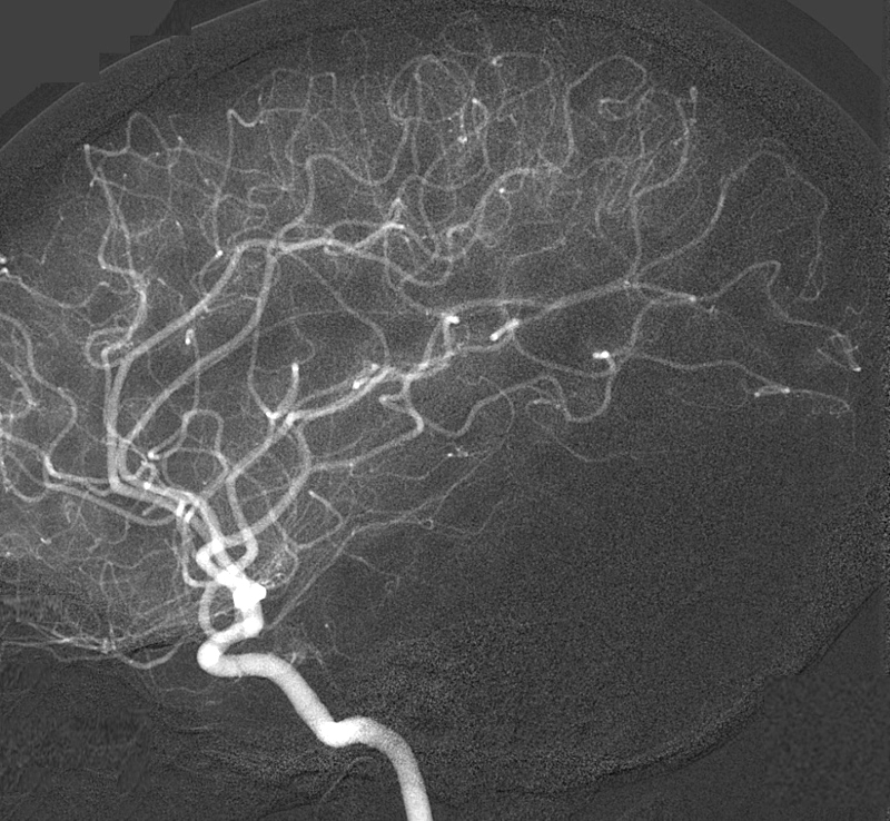
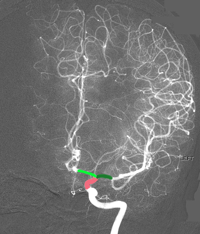
The Intracranial Circulation Derfinition of the Smaller Vessels Digital Angiogram of the Internal Carotid in the Lateral Projection |
|
The lateral digital angiogram of a selective internal carotid artery injection shows f the anterior and middle circulation supplied by the internal carotid artery. It is difficult to discern the origins of the anterior cerebral artery (ACA) and middle cerebral artery (MCA) in this projection. The study does however demonstrate the more distal portions of the ACA and MCA, and the smaller peripheral vessels. More specifically the loops of the distal middle cerebral arteries in the Sylvian fissure are well recognized. The intracranial portion of the internal carotid is well seen as well. Image Courtesy Ram Chavalli MD Copyright 2010 49420g01 |
|
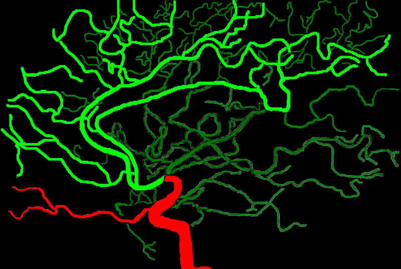
Anterior and Middle Cerebral Artery Distributions from a Lateral Perspective |
|
The anterior and middle cerebral circulation is demonstrated on the overlaid lateral angiogram of a selective internal carotid artery injection. The anterior cerebral circulation is overlaid in bright green and the middle cerebral circulation in dark green. The distal internal carotid artery and the ophthalmic artery (red) are shown (red) The ophthalmic artery is the smaller anterior horizontal vessel coursing to the orbit originates at the junction of the cavernous and supraclinoid segment. Image Courtesy of Philips Medical System Rendered by Ashley Davidoff MD Copyright 2010 All rights reserved 91390b01b04.8s |
|
The Intracranial Circulation Derfinition of the Smaller Vessels Digital Angiogram of the Internal Carotid in the A-P Projection |
|
The intracerebral angiogram shows the normal appearance in the A-P projection of the anterior and middle cerebral territory. The supraclinoid portion of the terminal portion of the internal carotid artery divides into the anterior cerebral artery (AC – bright green) that courses medially and the middle cerebral artery (MCA- dark green) that branches laterally. The study does also demonstrates the more distal portions of the ACA and MCA, and the smaller peripheral vessels. More specifically the loops of the distal middle cerebral arteries in the Sylvian fissure are well recognized. The supraclinoid portion of the internal carotid is overlaid in salmon pink. Image Courtesy of Ram Chavalli MD 49422g01 |
|
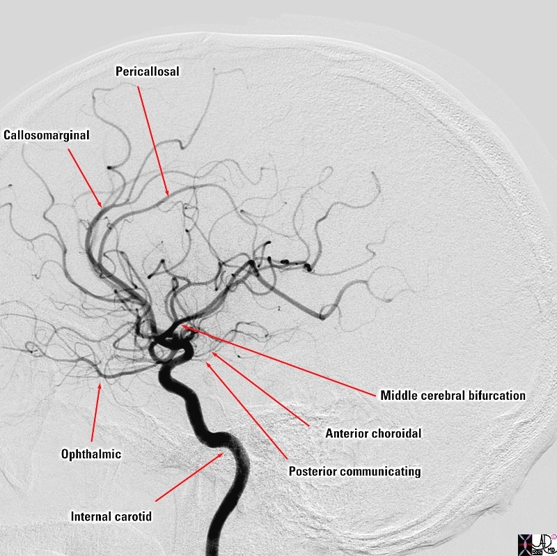
Intracranial Branches |
|
The selective of the internal carotid artery in the lateral projection reveals the major intracranial branches this vessel; The ophthalmic artery is is the first branch of the internal carotid artery distal to the cavernous sinus and it runs anteriorly to supply the eye, nose and meninges The ICA then gives off the posterior communicating artery that joins the circle of Willis followed by the anterior choroidal artery that supplies the choroid plexus but also many other forebrain structures including the optic chiasm, internal capsule, globus pallidus, tail of the caudate nucleus , amygdala. It also supplies midbrain structures including substantia nigra, red nucleus and crus cerebri After this it divides into a anterior cerebral artery that supplies the anterior and medial portion of the frontal lobes . It divides into the callosomarginal artery pericallosal artery middle cerebral artery that supplies the frontal region laterally, the parietal lobe, and the temporal lobe Courtesy Elisa Flower MD and Alex Norbash MD copyright 2010 97626b01.8s |
|
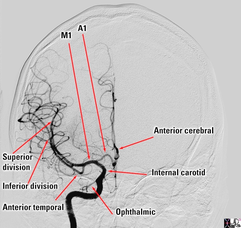
Intracranial Portion of the ICA – A-P Projection |
|
The selective of the internal carotid artery in the anteroposterior projection shows the major intracranial branches; The ophthalmic artery is is the first branch of the internal carotid artery distal to the cavernous sinus and it runs anteriorly to supply the eye, nose and meninges The ICA divides into the anterior cerebral artery (ACA) and the first segment is called the A1 segment that supplies the anterior and medial portion of the frontal lobes . middle cerebral artery (MCA) that supplies the frontal region laterally, the parietal lobe, and the temporal lobe Its first portion is called the M1 segment and it divides into two basic segments Superior division Inferior division The other vessel that can be seen in this view is the anterior temporal artery Courtesy Elisa Flower MD and Alex Norbash MD Copyright 2010 97628b.8s |
Course
Can be divided into five segments:
Segment A1 – before the anastomosis with the anterior communicating artery. It runs obliquely anteriorly and medially to the optic chiasm/nerve. In 30% of cases, it passes below the olfactory nerve. During its interhemispheric course, it runs mostly on the callosomarginal fissure.
It then heads to the interhemispheric fissure, finally anastomosing with its counterpart in the anterior communicating artery – Segment A2.
It gets in front of the lamina terminalis at the junction of the rostrum and genum of the corpus callosum.
The A3 segment contours the genum of the corpus callosum by a concavity directed backward.
The segments A4 and A5 travel posteriorly on the upper surface of the corpus callosum to the splenium.
Branches
The medial lenticulostriate arteries arise from segment A1 which irrigate the caudate nucleus and the anterior limb of the internal capsule
The recurrent artery of Heubner usually arises at the beginning of segment A2 irrigating the internal capsule. It lies along the basal segment and gives perforating branches in the anterior perforated space. It supplies the head of the caudate nucleus, globus pallidus and the anterior inferior part of the internal capsule
Orbitofrontal artery, arising from A2. It runs in the medial orbital sulcus, vascularizing the first two thirds of the orbital surface of frontal lobe (gyrus rectus and olfactory nerve).
The frontal polar artery also arises from A2. It heads upwards and forwards to the frontal pole, supplying the inner side of the anterior cingulate gyrus.
The pericallosal artery is actually the segment A3, entending posteriorly in the pericallosal sulcus to form the internal parietal arteries and precuneal artery. It gives several branches during its course in the cingulate sulcus.
Territory
The medial surface of the frontal lobe
The anterior four- fifths of the corpus callosum
Lateral surface of frontal and parietal lobe surrounding the medial longitudinal fissure
Anterior portions of the basal ganglia and internal capsule
Olfactory bulb and tract
Applied Biology
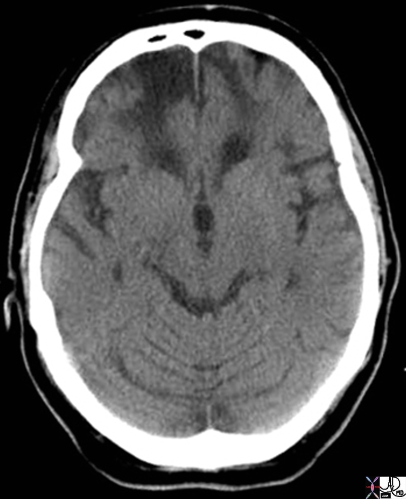
Chronic Bifrontal Infarcts |
|
The axial CT through the inferior aspect of the frontal lobes show blateral low density regions in the frontal lobes with volume loss indicative of chronic infarcts bilaterally with encephalomalacia Courtesy Ashley Davidoff MD Copyright 2010 71404 |
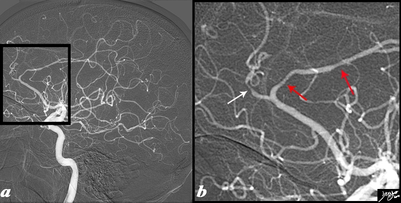
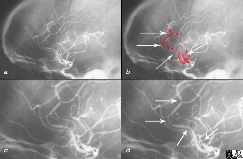
Vasospasm in the Anterior Cerebral Artery – Vasculitis |
|
The angiogram is from a 62 year old female patient is with acute neurological deficit The intracerebral angiogram shows focal regions of spasm with one area of spasm in the callosomarginal branch (white arrow) and two foci within the pericallosal artery (red arrows) . Image Ashley Davidoff MD Copyright 2010 89028c04.8s |
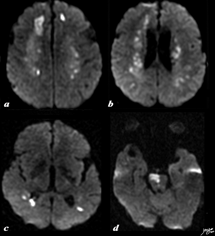
DWI – Multicentric Infarction – Acute Vasculitis |
|
The diffusion weighted MRI is from a 62 year old female patient is with acute neurological deficit The DWI sequence reveals multicentric of restricted diffusion indicating acute infarction. In image a, the dominant infarction is in the right frontal lobe, but there is an area of infarction in the left frontal lobe as well. Image b shows restricted diffusion in the periventricular white matter, while (c) shows disease in the posterior parietal region. In image (d) an infarction of the right side of the pons is apparent. Courtesy Ashley Davidoff MD Copyright 2010 89038c01.8s |
|
Adrenergic Induced Vasospasm in a Patient with Paraganglioma |
|
The angiogram is from a young woman who sustained a hemorrhagic stroke in the latterdays of her prgnancy> During the angiogram she had an “episode” of extreme anxiety and hypertension. The angiogram reveals multicentric foci of spasm in both the anterior and middle cerebral arteries. Workup following the hypertensive episode revealed a paraaortic paraganglioma. The diagnosis of contrast induced release of metanephrines from the paraganglioma was made which accounted for her reaction during the angiogram. The hypertension during her pregnancy accounted for her hemorrhagic stroke. Courtesy Ashley Davidoff MD 18450 |