The Common Vein Copyright 2010
Introduction
|
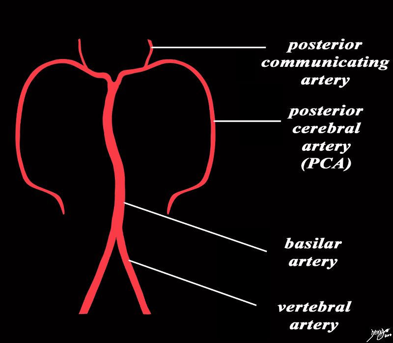
Basics of the Basilar Artery From Origin to Termination |
|
The diagram shows the main branches of the basilar artery. The basilar artery is formed by the two vertebral arteries and travel as a single artery over the upper medulla and the entire pons. Its terminal division is into the right and left posterior cerebral arteries. Its first branch after this division is the posterior communicating artery. Courtesy Ashley Davidoff MD copyright 2010 all rights reserved 97194b06b.8s |
|
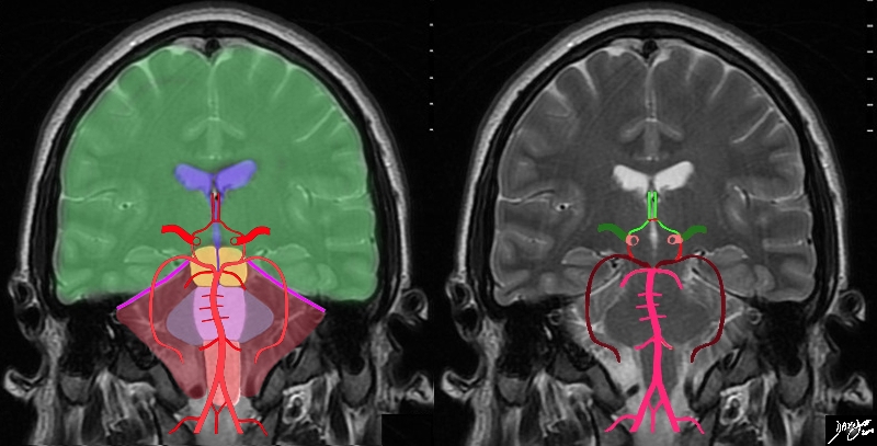
The Vertebral Arteries Part of the vertobasilar System |
|
The diagram shows the main branches of the blood supply to the brain including the circle of Willis overlaid on coronal MRI image to portray the approximate position of the vessels in the brain. The image on the right shows the combined system all in red, and the image on the right shows the derivation from the vertebrobasilar and carotid systems The carotid system supplies the brain from the internal carotid (salmon pink). We demonstrate its terminal bifurcation into middle cerebral (dark green) and anterior cerebral (bright green). The anterior communicating artery runs between the two anterior cerebrals (bright red) The basilar artery (pink) is formed by the two vertebral arteries and it travels as a single artery over the upper medulla and the pons. Its terminal branch is the posterior cerebral artery (maroon). The first branch off the posterior cerebrals is the posterior communicating which joins the middle cerebral to complete the circle of Willis Each of the carotid and vertebro-basilar systems contributes to the circle of Willis through communicating arteries. The vertebro-basilar system provides the posterior communicating arteries bilaterally from the posterior cerebral and the carotid system provides the anterior communicating arteries via the anterior cerebral arteries. Courtesy Ashley Davidoff MD Copyright 2010 All rights reserved 89721c06b.8sg05.8s |
|
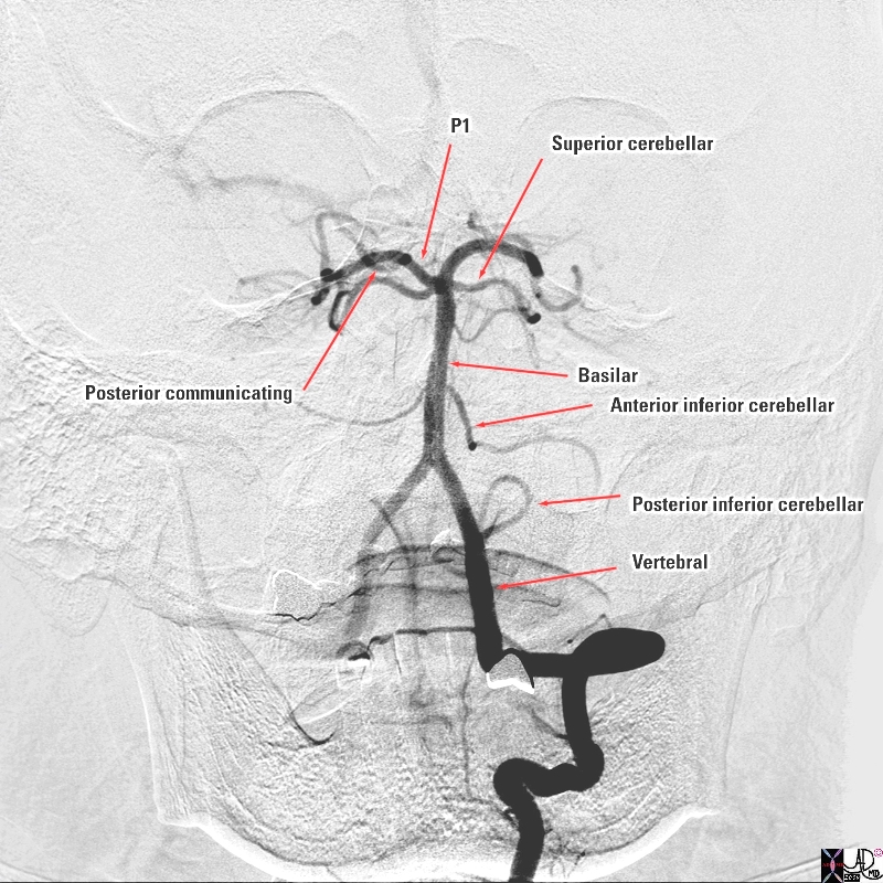
The Vertebral Artery and Major Branches Selective Angiogram in the A-P Projection |
|
The selective of the left vertebral artery in the A-P projection reveals the major intracranial branches; The posterior inferior cerebellar artery (PICA) which supplies the inferior aspect of the cerebellum The two vertebral arteries join to form the basilar artery which runs on the anterior surface of the pons. Its first branch is the; anterior inferior cerebellar artery ( AICA) which supplies the middle portion of the cerebellum and then the basilar artery gives off the superior cerebellar artery which supplies the superior aspect of the cerebellum The basilar artery finally terminates in the two posterior cerebral arteries (PCAs) which give off the posterior communicating arteries which connect with the circle of Willis and feed the inferior portion of the temporal lobes and the occipital lobes. Courtesy Elisa Flower MD and Alex Norbash MD Copyright 2010 97629b.8s |
|
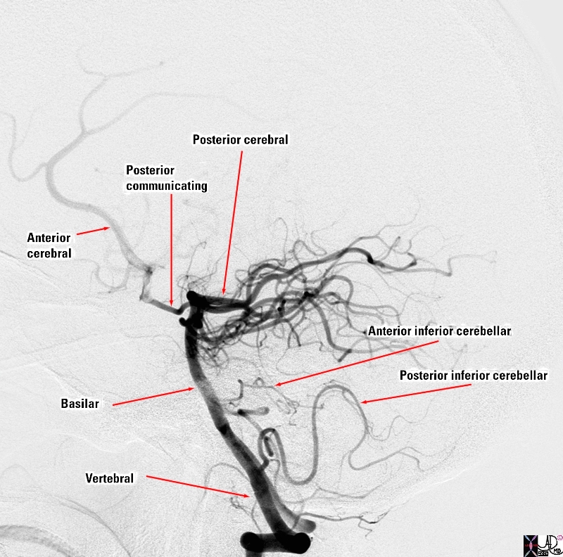
The Slective Angiogram in the Lateral Projection |
|
The selective angiogram of the vertebral artery in the lateral projection reveals the major intracranial branches; The posterior inferior cerebellar artery (PICA) which supplies the inferior aspect of the cerebellum The two vertebral arteries join to form the basilar artery which runs on the anterior surface of the pons. Its first branch is the; anterior inferior cerebellar artery ( AICA) which supplies the middle portion of the cerebellum and then the basilar artery gives off the superior cerebellar artery which supplies the superior aspect of the cerebellum (not easily distinguished in this view The basilar artery finally terminates in the two posterior cerebral arteries (PCAs) which give off the posterior communicating arteries which connect with the circle of Willis. The PCA feeds the inferior portion of the temporal lobes and the occipital lobes. The anterior cerebral artery is seen filling from the circle of Willis Courtesy Elisa Flower MD and Alex Norbash MD Copyright 2010 97630b02.8s |