
The Common Vein Copyright 2010
Introduction
From the Front |
|
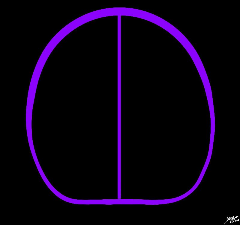
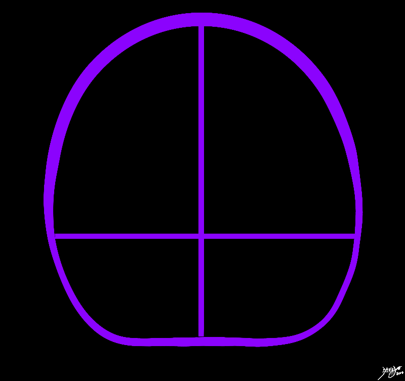
In the frontal or coronal view the brain has an almost rectangular shapeMidline structures and predominantly the interhemispheric fissure divides the brain into two sides Davidoff Art Copyright 2010 All rights reserved 93914b03.8s |
|
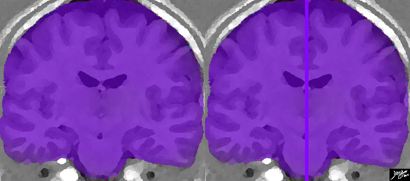
Frontal or Coronal View through the Middle of the Brain |
|
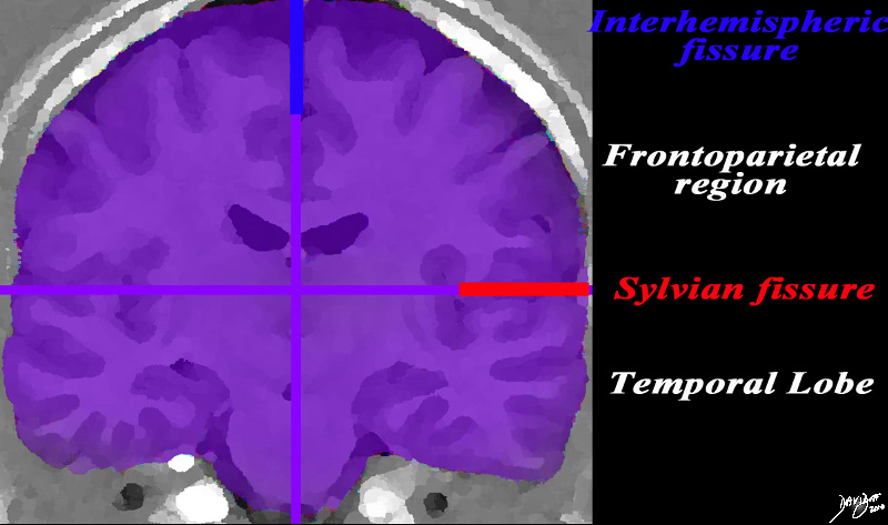
In the frontal or coronal view the brain has an almost rectangular shape, particulkalrly when viewed in a cut that traverses the temporal lobe. The midline structures of the brain, and predominantly the interhemispheric fissure divides the brain into two sides (purple divider). By convention, the cranial or superior part of the brain is above, the inferior part below with the right of the patient to your left and the left of the brain to your right. This rendered image of and MRI of the brain confirms the almost rectangular shape of the brain from this view and reveals the frontoparietal part of the brain in the anterior cranial fossa above, and the temporal lobes in the middle cranial fossa below. The internal anatomy will be discussed as we advance Davidoff Art Courtesy Ashley Davidoff MD Copyright 2010 All rights reserved 92167c01.8s |
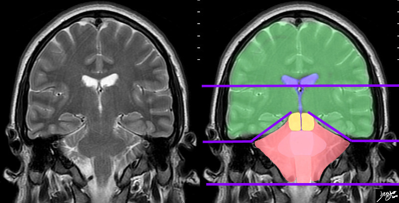
Forebrain Midbrain and Hindbrain |
|
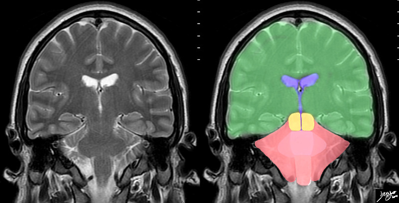
In this T2 weighted MRI image – the forebrain (green) midbrain (orange) and hindbrain (pink salmon containing only a small portion of the cerebellum have been outlined The ventricular system is outlined in blue Courtesy Ashley Davidoff MD copyright 2010 all rights reserve 89721c06b04.8s |
Midline Vertical Axis |
|
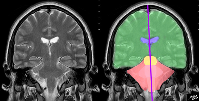
The vertical axis of the brain in the coronal plane is represented by many midline structures which in general are key relaying organs of the brain They include the ventricular ssytem, midbrain, pons, medulla and spinal cord Courtesy Ashley DAvidoff MD copyright 2010 89721c06b04.81s |
When the cut is taken anteriorly the anterior and middle cranial fossa are exposed, but if it is taken posteriorly the anterior and posterior cranial fossae are exposed.
|
Anteriorly Space for the Frontal and Parietal Lobes above and the Temporal Lobes below |
|
Anteriorly the anterior and middle cranial fossae are apparent. Midline structures and predominantly the interhemispheric fissure divides the brain into two sides Davidoff Art Copyright 2010 All rights reserved 93914b04.8s |
|
Important Fissural Landmarks in the Coronal Plane |
|
In the same cut the fissural landmarks enable us to divide the brain into the two hemispheres (blue interhemispheric fissure) and the Sylvian fissure (red) divides the frontoparietal region in the anterior craial fossa from the temporal lobe in the middle cranial fossa. Davidoff Art Courtesy Ashley Davidoff MD Copyright 2010 All rights reserved 92167..84s label |
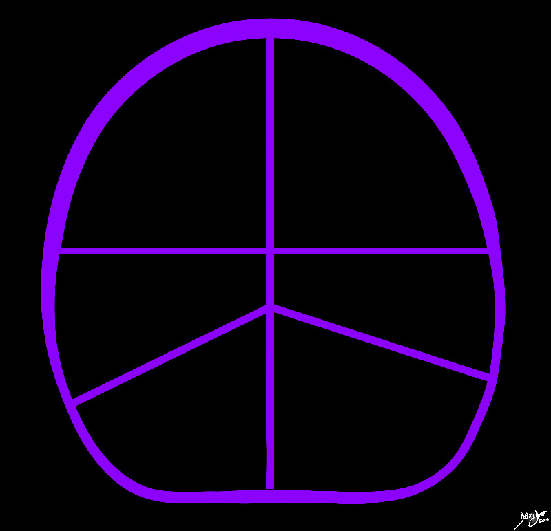
The Three Levels of the Cranium |
|
The drawing reveals the three levels of the brain in the coronal plane corresponding in general to the anterior cranial fossa (upper level), middle cranial fossa (middle level) and posterior cranial fossa (lower level) Courtesy Ashley Davidoff MD Copyright 2010 all rights reserved 93914b05b.8s |
The Horizontal lines and a Rough Guide to the Cranial Fossae |
|
The horizontal axes divides the brain into the fossae in which the various parts of the brain reside. The top story is the anterior cranial fossa where most of the forebrain resides, the middle story is the middle cranial fossa where ythe temporal lobes and occipital lobes , and the ground floor is the posterior cranial fossa where the hindbrain resides Courtesy Ashley Davidoff MD copyright 2010 89721c06b04.85s |
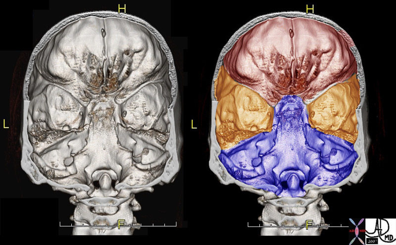
The Cranial Fossae |
| 48720c01 brain cerebrum cerebral skull anterior cranial fossa middle cranial fossa posterior cranial fossa space position protection bone skull anatomy applied biology TCV the common vein CTscan 3D volume rendering Davidoff MD |