
The Common Vein Copyright 2010
Introduction
|
The vectors of the ventricular System Overlaid on the Brain |
|
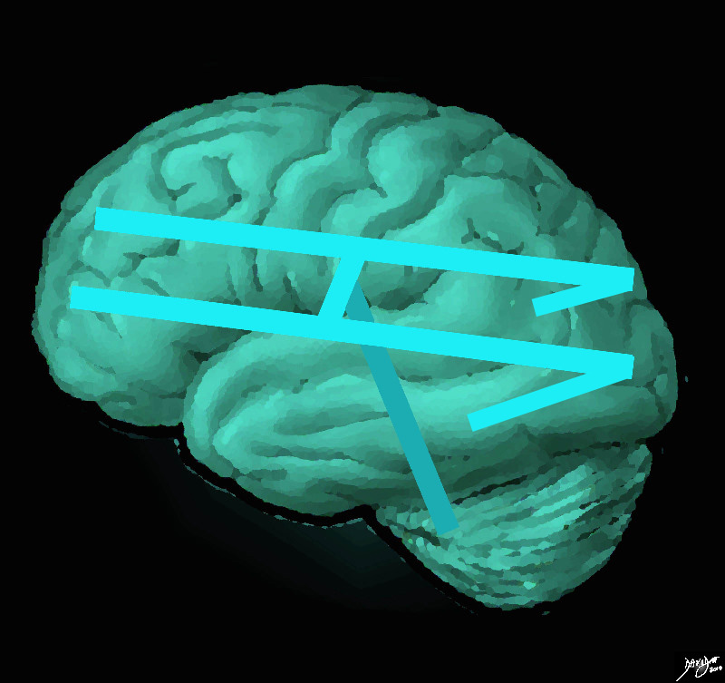
The ventricular system is overlaid on a drawing of the brain. Each limb of the horizontal component is situated in a cerebral hemisphere. The angled parts that extend inferiorly represent the temporal horns. The vertical limb includes the third and fourth ventricles Courtesy Ashley Davidoff MD copyright 2010 all rights reserved 94458e07.81s |
 The Deepest Layers of the Inverted C’s The Deepest Layers of the Inverted C’s |
|
In the diagram that describes the series of inverted “C” the ventricles are the deepest and most midline of the structures In this diagram it is demonstrated as the innermost teal bluue ring. The frontal horns are superior and anterior and the temporal horns are the most inferior and anterior. Courtesy Ashley Davidoff MD copyright 2010 all rights reserved 93890b01b05b1.8s |
|
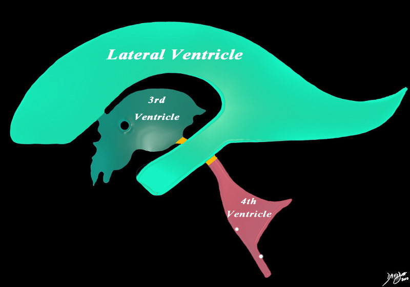
The Major Components of the Ventricles |
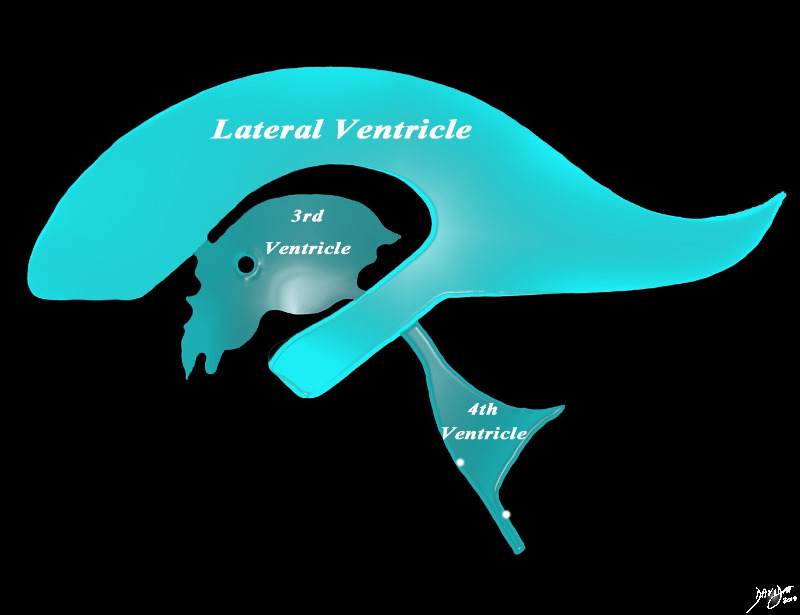
| The diagram in the sagital projection reveals the horizontal portion called the lateral ventricle. It is a paired structure.The vertical portion (light green consists of the midline 3rd and 4th ventricle.Courtesy Ashley Davidoff MD copyright 2010 all rights reserved 94459b06b.8s |
|
Detailed Components |
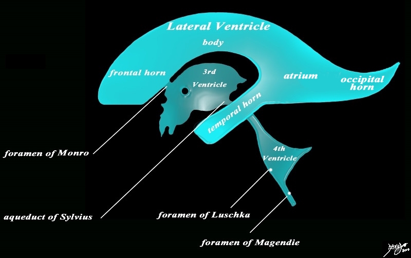
| The diagram in the sagital projection reveals the horizontal portion called the lateral ventricle. It is a paired structure, and houses the frontal horn, body, occipital horn and atrium. The vertical portion consists of the paired foramina of Munro, the midline third ventricle, the narro aqueduct of Sylvius, the 4th ventricle the anteriorly placed paired foramina of Luschka, and theposteriorly positioned foramenCourtesy Ashley Davidoff MD copyright 2010 all rights reserved 94459b10b02.82s |
|
The Recesses |
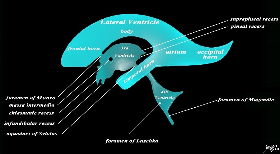
| The sagital projection reveals the horizontal portion called the lateral ventricle. It is a paired structure, and houses the frontal horn, body, occipital horn and atrium. The vertical portion consists of the paired foramina of Munro, the midline third ventricle, the narrow aqueduct of Sylvius, the 4th ventricle the anteriorly placed paired foramina of Luschka, and the posteriorly positioned foramen of Magendie There are 4 recesses that create an unusual shape to the third ventricle. Anteriorly the chiasmatic (optic) recesslies above the the infundibular recess. Posteriorly the sprapineal recess lies above the pineal recess.Courtesy Ashley DAvidoff MD copyright 2010 all rights reserved 94459b14b.82s |
Distribution Bewteen Forebrain Midbrain and Hindbrain
 Distribution of the Ventricles Distribution of the Ventricles |
|
The diagram in the sagital projection reveals the anatomical distribution of the ventriclular components into the forebrain (green), midbrain (orange) and hindbrain (salmon). Courtesy Ashley DAvidoff MD copyright 2010 all rights reserved 94459b06b01.81s |