The Common Vein Copyright 2010
Deinition
Anterior Limb of Internal Capsule
White matter structure that separates the caudate nucleus and the thalamus from the lenticular nucleus. The anterior fibers extend from the frontal cortex to the pons as well as connect medial and anterior thalamic nuclei to the frontal lobes. Dysfunction leads to contrahemisperic sensory and motor function loss as well as problems with vision.
Posterior Limb of Internal Capsule
The posterior fibers consist of corticospinal and sensory-somatic fibers.
|
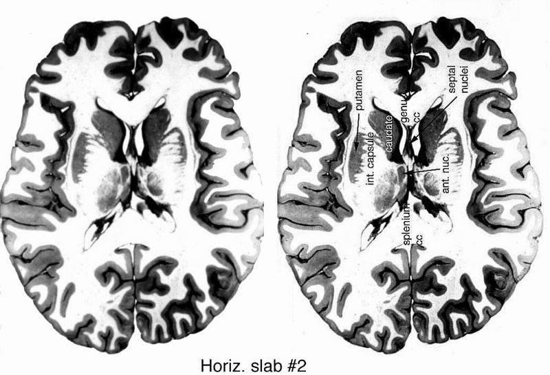
The Internal Capsulein Transverse Section Anatomic Specimen |
|
The anatomic section is an axial slice through the brain showing the ventricles in midline followed by the genu of the corpus callosum anteriorly and splenium posteriorly, the caudate nucleus, the anterior nucleus, putamen and then surrounded by white matter followed by the gray matter Courtesy Courtesy Department of Anatomy and Neurobiology at Boston University School of Medicine Dr. Jennifer Luebke, and Dr. Douglas Rosene 97135c.8 |
|
Putamen in Cross Section T2 Weighted MRI |
|
The axial T2 weighted MRI is taken through the level of the ventricles. It demonstrates the midline ventricuar system, the and the orange ring of paraventricuar basal gaglia including tye caudate nuclii (most anterior and medial) the and the putaen. It also demonstrates putamen which is a pink slmomn in color lying lateral to the white matter. The thalami ar noted posteriorly. The body of the corpus callosum is Courtesy Ashley Davidoff MD copyright 2010 all rights reserved 94079cd07b.81sh |

|
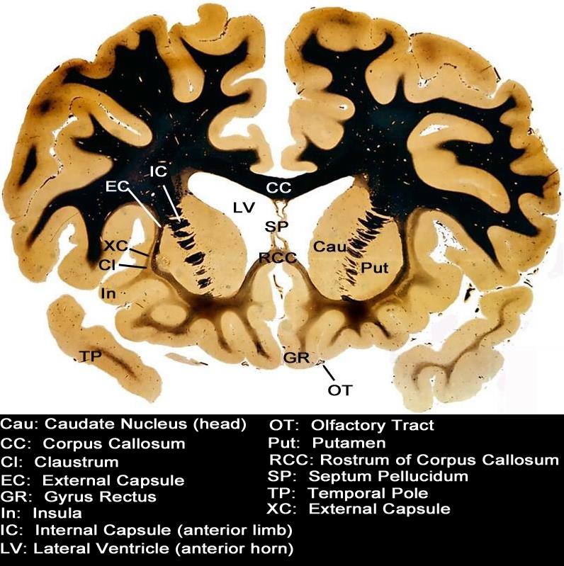
Coronal Section |
|
This coronal section through the forebrain reveals the more superficial cortical structures that include the frontoparietal region and the temporal pole(TP). Centrally the corpus callosum (CC) and septum pellucidum SP are noted, and are surrounded by the next paracentral structures which include the left and right lateral ventricles (LV). The basal ganglia starting with the head of the caudate nucleus (Cau) and then the putamen (Put) follow, and these basal ganglia are separated by the internal capsule (IC). The external capsule (XC) and claustrum (Cl) follow as the two lateral structures till we return to the outer umbrella of the frontoparietal cortex again. Courtesy Department of Anatomy and Neurobiology at Boston University School of Medicine Dr. Jennifer Luebke, and Dr. Douglas Rosene 97342.C3.8L01 |