
Embryology
Copyright 2009
Author
Introduction
The central nervous system arises from the ectoderm as the neural plate. The neural plate folds to form a neural tube.
Week 1-3
Day 18 (crown rump length CRL = 1.5mms)
The neural plate begins to thicken at the margins and the neural groove begins to form
Day 19
The prosencephalon, mesencephalon and the rhomboencephalon, are differentiated by small neural folds.Day 21 (CRL = 3mms)
optic vesicles formed
Day 22
The folding of the neural tube begins
Week 3-4
Day 25
At about day 25 after closure of the neural tube, sthere is subdivision and expansion of the neural tube with the prosencephalon mesencephalon and rhombencephalon becoming more distinct.
|
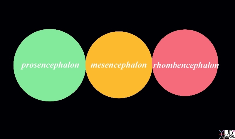
About 4th Week 3 primary Fluid Filled Vesicles Prosencephalon Mesencephalomn and Rhombencepahalon |
|
At about 4th week when the when the crown rump length is about 3mm, the prosencephalon, mesencephalon and the rhombencephalon, are differentiated by small neural folds. Davidoff Art Courtesy Ashley Davidoff MD Copyright 2010 98481b.8L |
Day 26 (CRL = 3.00 mms)
The neural canal communicates with the amniotic fluid via the cranial and caudal openings or neuropores which close at about this time.
Rapid growth causes the neural tube to fold ventrally
Day 27 (CRL = 3.3mms)
closure of posterior neuropore
ventral horns appear
Brain and head represent 50% of total body length
Week 5
Day 31 (CRL = 4.3mms )
At this stage during week 5 there is division of the prosencephalon and rhombencephalon, each, into two separate or secondary vesicles. The prosencephalon divides into an anterior telencephalon and a posterior diencephalon. The rhombencephalon divides into the metencephalon (future cerebellum and pons) and myelencephalon (future medulla).
The lateral ventricles form within the telencephalon, and the central lumen of the diencephalon will develop into the the third ventricle. The aqueduct of Sylvius is formed from within the mesencephalon and the fourth ventricle is frormed from within the rhomboencephalon.
It is during this time also, at about the fourth week that the primitive nervous system starts to fold. There are a few bends or flexures – The cranial flexure results in the prosencephalon flexing ventrally. The midbrain flexure is the most prominent flexure and develops along the ventral aspect of the mesencephalon.The cervical flexure develops at the junction of the myelencephalon and spinal cord.. There is also a pontine flexure between the midbrain and rhombencephalon.
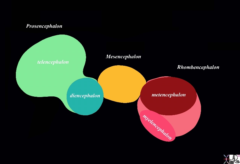
5 Vesicles and Flexures Formed During Rapid Growth and Differentiation |
|
At about day 31 or 32 there is division of the prosencephalon and rhombencephalon, each, into two separate or secondary vesicles. The prosencephalon divides into an anterior telencephalon and a posterior diencephalon. The rhombencephalon divides into the metencephalon and myelencephalon. There are now 5 vesicles making up the primitive brain Note also that the rapid growth of the brain forces it to start to bend forward and ventrally Courtesy Ashley Davidoff MD Copyright 2010 98481b02b01.8s |
Day 35 (CRL = 5.0mms)
5 cerebral vesicles are recognised
Prosencephalon (telencephalon diencephalon)
Mesencephalon
Rhombencephalon (metencephalon and myelencephalon)
Week 5
In each of the 5 vesicles of the neural tube, the central lumen develops into the ventricular system .
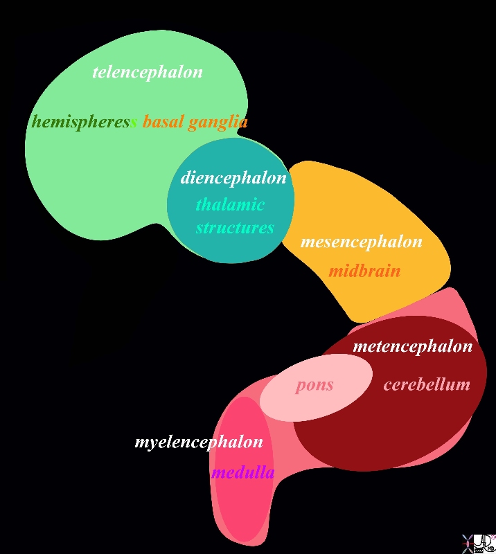
Further Flexion and Bending With Rapid Growth |
|
With further growth the flexures result in further ventral and horizontal positioning of the forebrain in relation to the mid and hindbrain This diagram outlines the alternative terminology for the developing brain. Courtesy Ashley Davidoff MD Copyright 2010 98481b03b05.8s |
Week 6-8
Ears develop
Limbs develop
Day 42 (CRL = 13mms )
primordium cerebellum
Day 56 (CRL = 25mms)
differentiation of cerebral cortex and meninges
Day 150 (CRL = 225mms)
primary cerebral fissures appear
Day 180 (CRL = 230mms)
Secondary cerebral sulci
Weeks 13-20
Rapid brain growth
Myelination begins
Weeks 21-40
Birth
Head 25% of body length
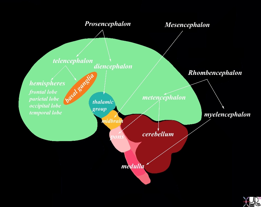
The Final Product – Ready to Take on the World |
|
With continued growth and especially growth of the forebrain and more specifically of the cerebral hemispheres the shape of the brain takes on its distinctive form as depicted in this image in the sagittal plane. This diagram again outlines the alternative terminology for the developing brain. Courtesy Ashley Davidoff MD Copyright 2010 98481b06b01.9s |
References
Bionalogy.com – Excellent Graphics