
Hydrocephalus
The Common Vein Copyright 2010
Definition
Hydrocephalus is a progressive distension of the ventricles, caused by an abnormality of either the production of cerebrospinal fluid or its circulation/resorption.
The clinical features and prognosis depend primarily on age. When the sutures of the skull are still unclosed, the result is a progressive macrocephaly. When sutures are closed in older children (after 20 months), hydrocephalus results in a syndrome of intracranial hypertension.
Multiple mechanisms are identified; there can be hydrocephalus from obstacles at the interventricular foramen, at the aqueduct of Sylvius, at the foramina of Magendie and Luschka or at the subarachnoid space (sequelae of arachnoiditis). There can also be an excessive production from choroid processes, and decreased CSF resorption at the granulations Pacchioni (altered granulations, or venous hypertension in the superior sagittal sinus).
It can be associated with cranial/cerebral malformations, meningitis, cerebral hemorrhage, tumors.
Clinically, a progressive macrocephaly is observed, which is why the measurement of head circumference is routinely done in pediatric age. There is also a bulging fontanel, scalp skin thin, tense, with venous dilatation.
It can be detected during pregnancy if the mechanism is present during the fetal stage by ultrasound as early as the 16th week of gestation, for which a confirmation is done between the 20th and 22nd week.
The treatment of hydrocephalus is mainly neurosurgical.
Giant Cell Astrocytoma with Secondary Hydrocephalus |
|
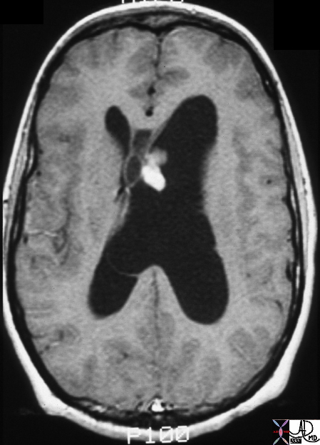
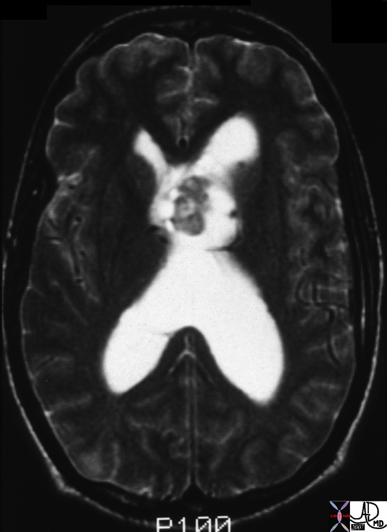
A mass close to the 3rd ventricle and septum pellucidum displaces and compresses the right lateral ventricle and results in hydrocephalus seen as dilted butterfly shape (black on T1) and white on T2 Courtesy James Donnelly MD 23063 23064 |
Central Neurocytoma |
|
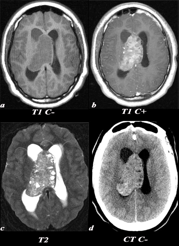
A 21 year old female was found to have papilledema on physical exam noted by her optometrist. MRI: T1 pre (T1 C- a) and post (T1 C+ b): Post contrast images demonstrate the heterogeneous enhancing nature of this mass. These images demonstrate centered in the right lateral ventricle with involvement of the septum pellucidum and partial extension into the left lateral ventricle. T2: T2 weighted images demonstrate internal areas of high signal consistent with cystic components. Also note the high T2 signal adjacent to the enlarged lateral ventricles which is a finding consistent with hydrocephalus, classically called transependymal flow of CSF. This unenhanced CT scan demonstrates also demonstrates the mass in the right lateral ventricle with similar features of heterogeneity and involvement of the septum pellucidum and partial extension into the left lateral ventricle. Notice the internal low density or cystic components and the higher density calcifications seen posteriorly. The lateral ventricles are enlarged consistent with resultant hydrocephalus. These findings are consistent with a diagnosis of central neurocytoma. Image Courtesy Elisa Flower MD and Asim Mian MD 97634c01.8s |
Desmoplastic Infantile Ganglioglioma Compressing the Third Ventricle |
|
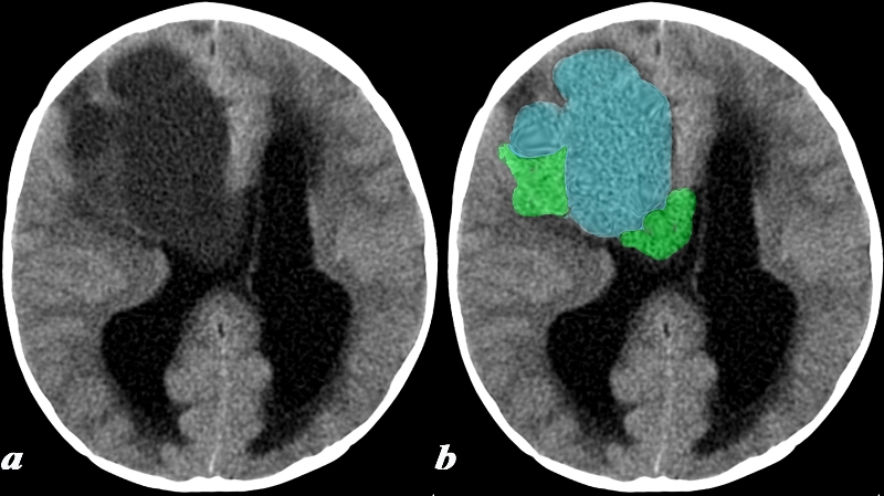
This 17 month old boy presented with vomiting. CT: (a,b) A noncontrast head CT was obtained and demonstrates a predominantly cystic lesion (blue) with more solid components (green) at the periphery. Notice the enlargement of the lateral ventricles which is due to obstructive hydrocephalus caused by obstruction at the level of the third ventricle (not included on this image). These findings are consistent with a desmoplastic infantile ganglioglioma (DIG) Image Courtesy Elisa Flower MD and Asim Mian MD 97641c.8 |
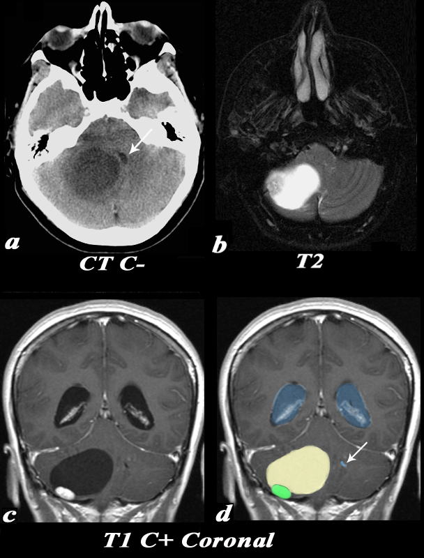
hemangioblastoma Compressing the 4th Ventricle – Hydrocephalus |
|
This 55 year old female complained of an occipital headache for one month with increased difficulty ambulating. CT: Noncontrast CT image (a) demonstrates a large low density lesion in the right cerebellum. This lesion causes significant mass effect on the fourth ventricle which is displaced towards the left (white arrow). This mass effect caused obstruction of the CSF pathway resulting in obstructive hydrocephalus and subsequent symptoms. MRI: The T2 weighted image (b) demonstrates a predominantly T2 hyperintense cystic lesion with a nodular T2 isointense component seen laterally. Post contrast T1 weighted coronal images (identified by high signal in the venous sinuses demonstrate the classic cyst (light yellow in d) with mural nodule (green in d) appearance of a hemangioblastoma. Note the enlarged lateral ventricles (blue) and the displaced and compressed 4th ventricle (white arrow d). Image Courtesy Elisa Flower MD and Asim Mian MD 97652c01.81 |