Results
The Common Vein Copyright 2010
Introduction
|
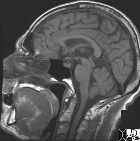
T1 Weighted Sagittal Image Normal Brain |
|
The T1 weighted images have good spatial resolution but inferior contrast resolution meaning in the latter instance that the images are more gray when compared to more black and white contrats of T2 weighted images. Fat though is quite bright as seen in the subcutaneous tissues and to lesser extent in the bone marrow. Air in the maxillary sinus is quite black while the CSF is a dull charcoal. White matter like the corpus callosum are well seen and the midbrain hind brain, and spinal cord are quite well seen. 49079b01 brain cerebrum cerebral normal anatomy MRI T1 weighted sagital Davidoff MD |
|
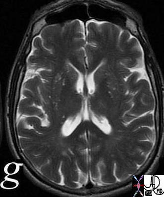
T2 Weighted Axial Image Through the Middle of the Lateral Ventricles |
|
This axial image of a normal T2 weighted MRI scan shows high intensity CSF anatomy. Note the presence of the lower signal intensity of the choroid plexus in the occipital horms and the excquisite definirtion of the thin sulci and hence the gyral pattern. In this instance the T2 characteristics of the the caudate nuclii that lie just lateral and posterior to the frontal horns are very similar to the putamen which also retains slight hyperintensity with tiny punctate ares of T2 hyperintensity. The corpus callosum on the other hand including the “V” shaped genu between the frontal horns, and the inverted V shaped splenium between the posterior occipital horns are composed of white matter and contain little water and are therefore quite dark on the T2 weighted images. Courtesy Ashley Davidoff MD 38703c03g code CNS brain CSF normal |