External Carotid Artery
Copyright 2009
Definition
need to editSection Text
“The external carotid artery springs from the common carotid artery opposite the top of the thyroid cartilage, and from there it courses up, curves forward and then back to an area behind the neck of the mandible. The artery diminishes in size along its route, and is approxiamtely equal in size to the internal carotid artery. Near to the external carotid artery at the center of the body is the hyoid bone, as well as the pharymx’s wall, the superior laryngeal nerve, and some of the paroitd gland. Near to it away from teh body’s center is the internal carotid artery, while is the superior laryngeal nerve. Higher up, the external carotid artery is separated from the internal carotid artery by the styloglossus, the stylopharyngeus, the glossopharyngeal nerve, the pharyngeal branch of the vagus nerve, and some of the parotid gland. It is covered by the skn, subcutaneous fascia, platysma, deep fascia, amd the anterior margin of the sternocleidomastoideus. The vessel is crossed by the hypoglossal nerve, the lingual vein, the ranine vein, the common facial vein, the superior thyroid vein, the digastricus, and the stylohyoideus. The external carotid artery’s branches, in order of origin, are the superior thyroid artery, the ascending pharyngeal artery, the lingual artery, the facial artery, the occipital artery, the posterior auricular artery, the superifical temporal artery, and the maxillary artery.”
|
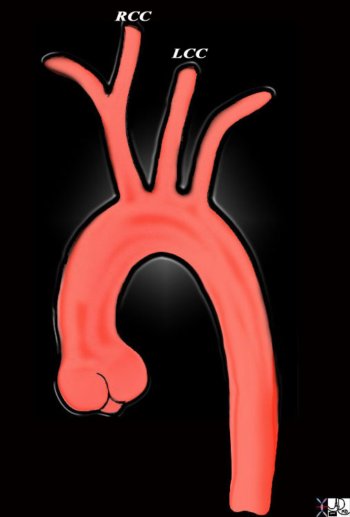
Origin of the Carotids |
|
This diagram shows the origin of the head neck and arm vessels from the arch of the aorta. The first vessel off the arch is the brachiocephalic or innominate artery and it branches into the right common carotid (RCC) and right subclavian artery. The second vessel is the left common carotid artery (LCC) and this is followed by the left subclavian artery. The RCC and LCC divide in the neck into the external and internal carotid vessels 47676b Courtesy Ashley Davidoff MD Copyright 2010 |
|
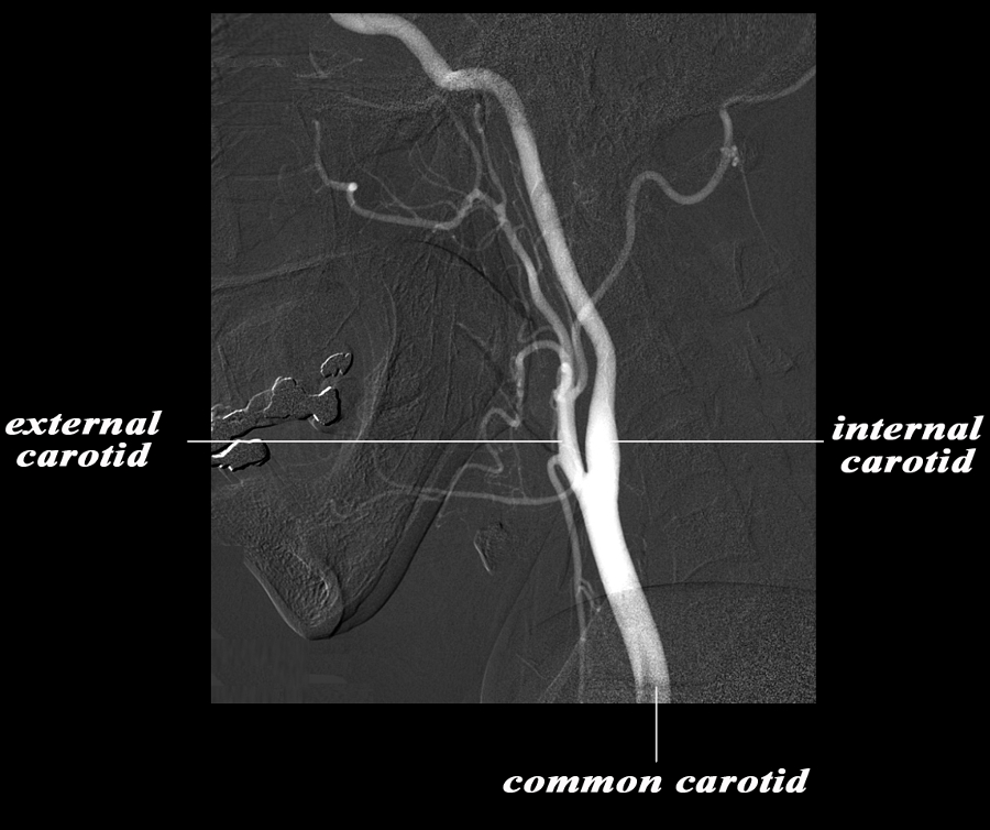
The Common Carotid and Terminal Branches |
|
The angiogram demonstrates the carotid system in the lateral projection. The common carotid artery bifurcates in the base of the neck into the external and internal carotid artery. Basically the internal carotid artery supplies the brain and the external supplies the structures of the face and muscles and skin of the cranium. The carotid system supplies the brain from the internal carotid – a branch of the common carotid which arises from the aorta. Courtesy Ram Chavalli MD 49400bc01.8s |
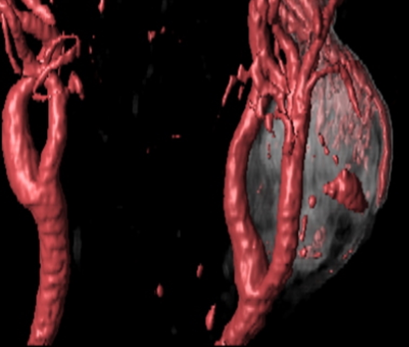
Carotid Body Tumor |
|
The CTA with volume rendering shows a mass (gray) at the bifurcation of the left common carotid. This location is classical for a carotid body tumor – or chemodactoma Courtesy Philips Medical Systems 92506.8 |
|
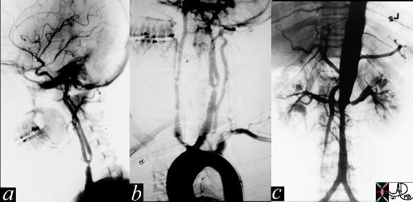
Takayasu’s Arteritis |
|
The series of images are from the angiogram of a 14 year old female who presented with seizures and an elevated blood pressure. Images a and b show multiple stenoses within the carotids best seen at the level of the bifurcation into external and internal arteries. In addition in b, the aortic arch shows non critical narrowing just after the origin of the left common carotid vessel. Note that the right subclavian artery is not seen and presumably is accluded at its origin. The abdominal angiogram shows a significant narrowing of the left renal artery with post stenotic dilitation, and stenotic disease in the infrarenal abdominal aorta. The multicentric nature of the disease in a young female is athognomonic of Takayasu’s arteritis. 35155c Courtesy of Laura Feldman MD. |