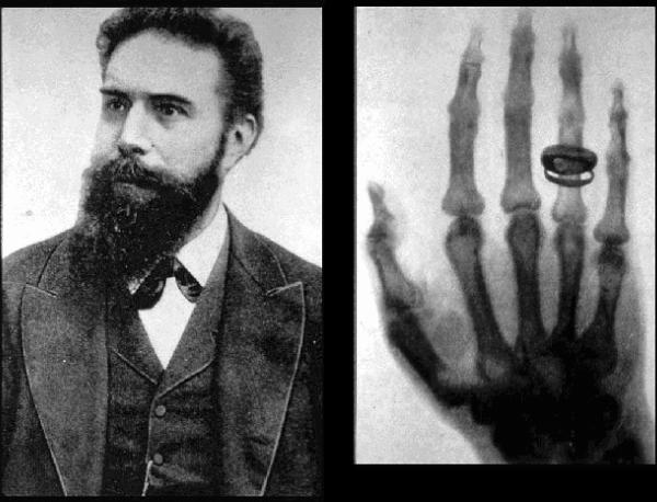
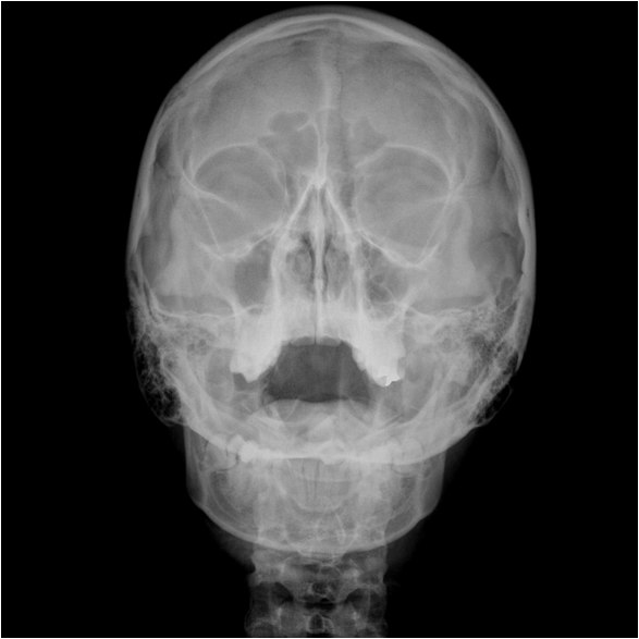
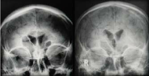
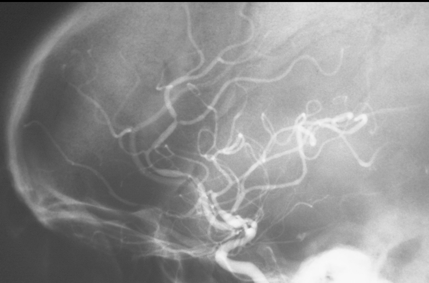
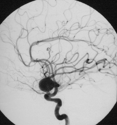
Roentgen with a historic image of his wife’s hand and wedding band. November 8, 1895: Roentgen’s Discovery of X-Rays 54463 #historicalWaters view showing diffuse prominent mucosal thickening in the right maxillary sinus and mild nmucosal thickening in the left maxillary sinus. Please note that normal aeration in the maxillary sinuses is demonstrated as relative decreased density of the maxillary sinuses when compared to the density of the orbita. Courtesy Erhan Erdogan, WikimediaLumbar air encephalogram from Annual Oration More Than The Sum Of Its Parts Royal Victoria Hospital, Thursday 4th October 2007 Stanley A Hawkins The Ulster medical journal · February 2008Conventional Angiography Showing Multifocal Regions of Vasospasm Ashley Davidoff TheCommonVein.netDigital Angiography showing a Large Berry Aneurysm Ashley Davidoff TheCommonVein.net
CT scan
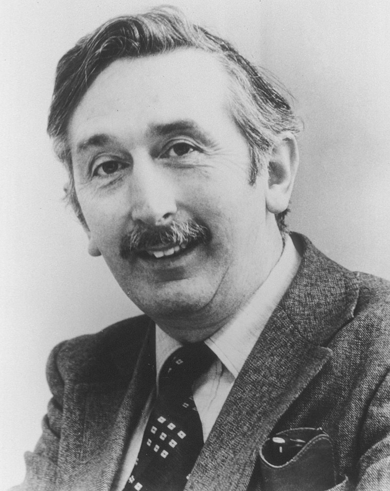
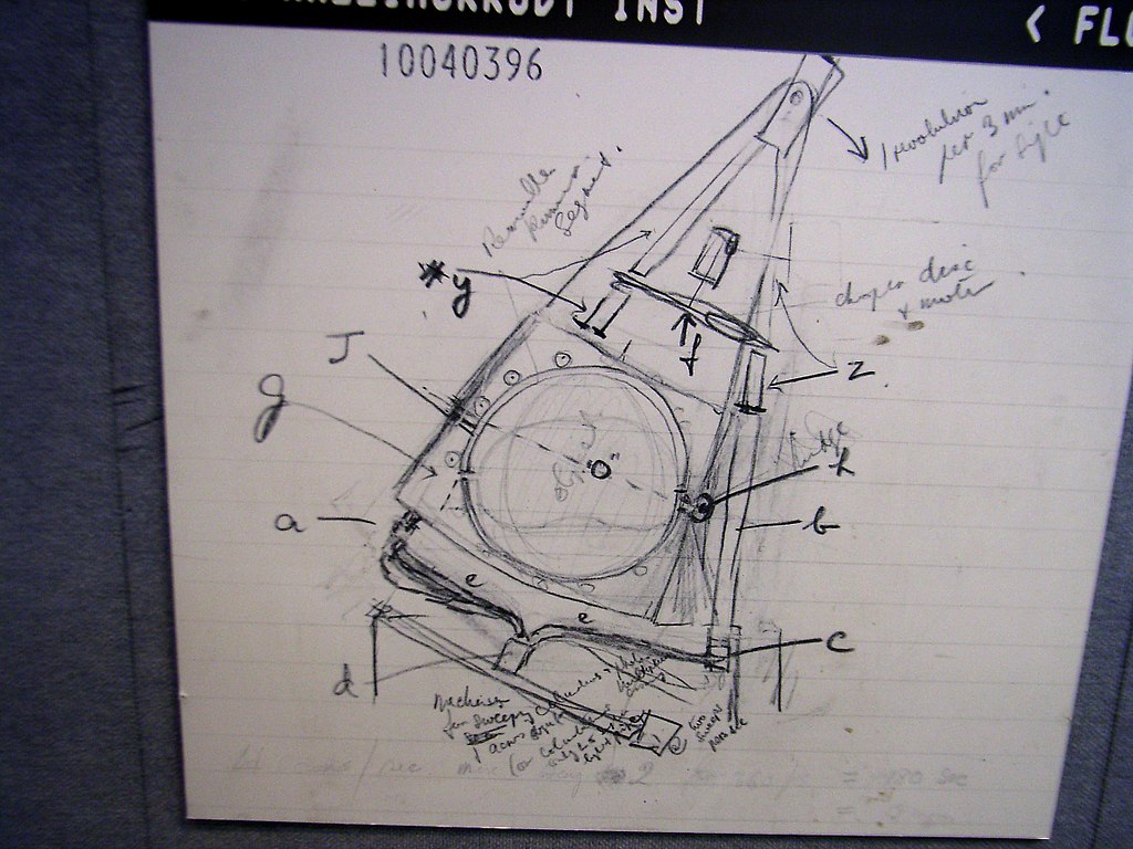
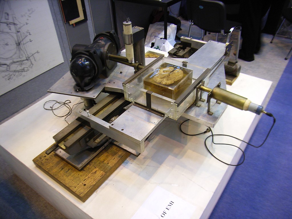
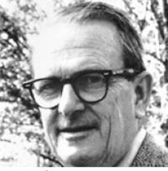
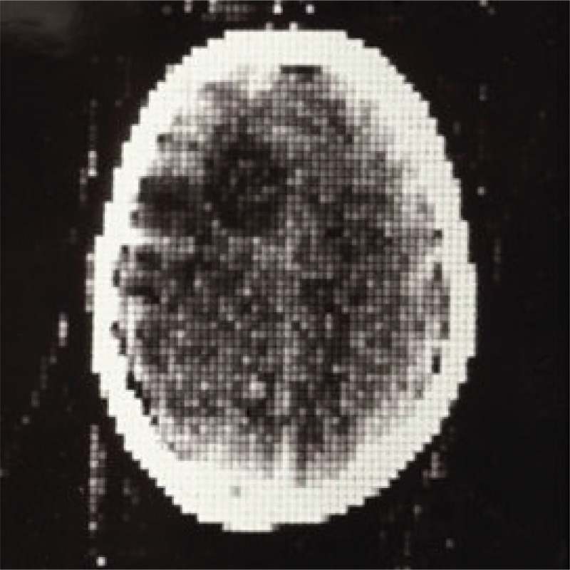
Godfrey N. Hounsfield 1975 Nobel prize Award Winner –Hounsfield’s sketch of the prototype CT scanner. Original sketch from Hounsfield’s notebook. The picture was taken at the UKRC 2005 exhibition, Manchester G-Mex centre. 08:56, 20 July 2006 (UTC) WikiThe very first CT scanner prototype. Invented by Hounsfield at EMI. This picture was taken at the UKRC 2005 exhibition in Manchester G-MEX centre. Philipcosson 08:42, 20 July 2006 WikiExample of the Image ObtainedAllan Macleod Cormack South African American physicist who won the 1979 Nobel Prize in Physiology or Medicine (along with Godfrey Hounsfield) for his work on X-ray computed tomography (CT) WikiThe first clinical CT scan, acquired October 1971 at Atkinson Morley’s Hospital in London
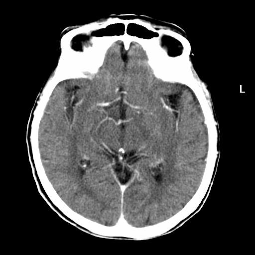
Normal CT
Normal CT scan of the Brain with Contrast showing the Circle of Willis Ashley Davidoff TheCommonVein.net
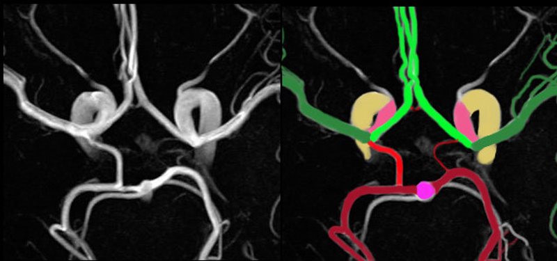
CTA
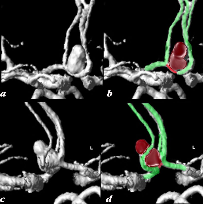
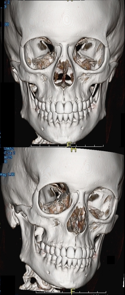
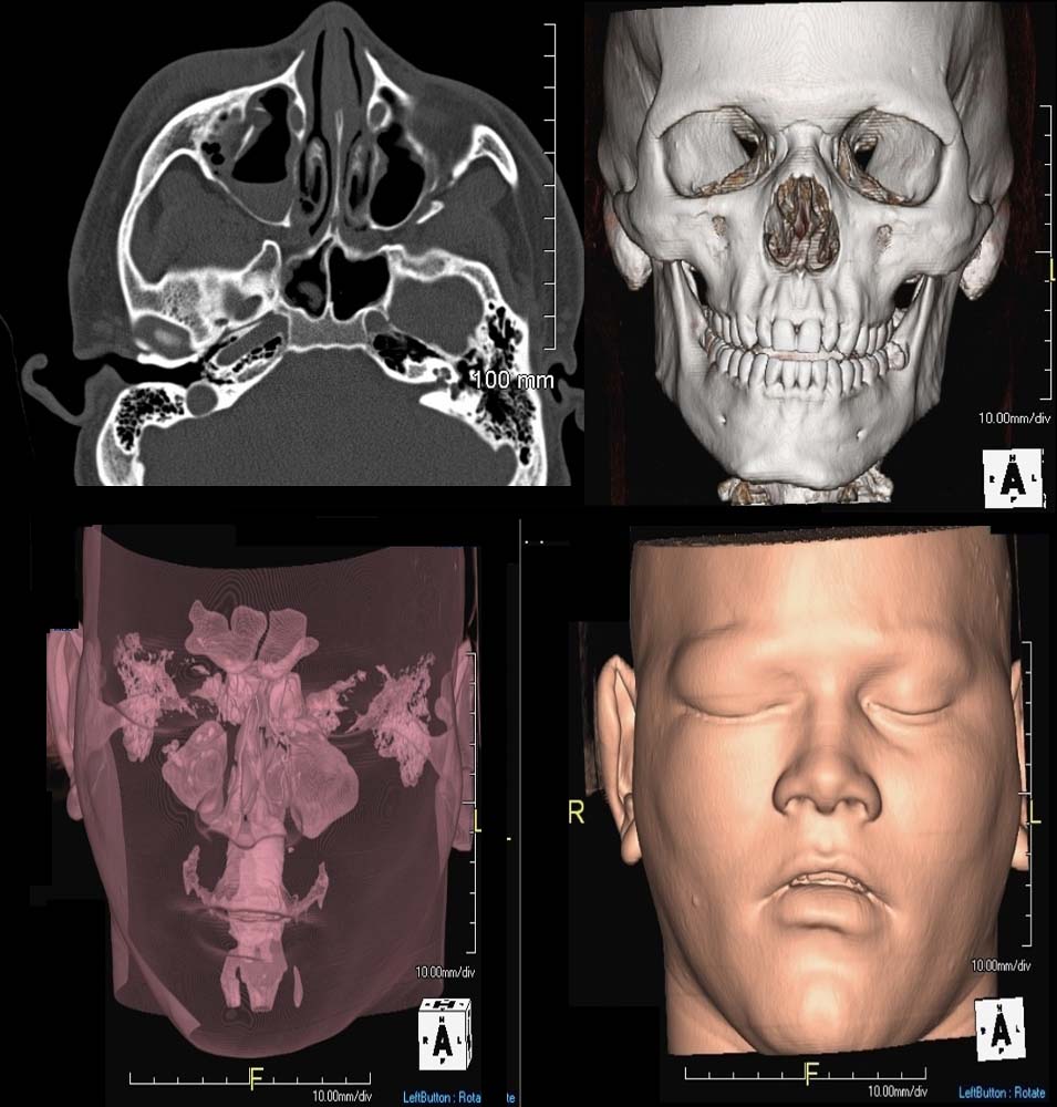
The axial MRA using 3T magnet shows a magnified view of the circle of Willis overlaid in color to reflect the component vessels. The supraclinoid portion of the internal carotid (salmon pink) divides into the middle cerebral (MCA dark green) and anterior cerebral (ACA bright green). There is a tiny anterior communicating artery (bright red) that runs between the two anterior cerebrals. The basilar artery (bright pink) gives rise to the posterior cerebral (PCA maroon) and superior cerebellar arteries (white) carotid system. The posterior cerebral artery gives rise to the posterior communicating artery which plugs into the region of the origins of the MCA and ACA. Note the left posterior communicating artery is relatively small which is a normal variant. Each of the carotid and vertebro-basilar systems contributes to the circle of Willis through communicating arteries. The vertebro-basilar system provides the posterior communicating arteries bilaterally from the posterior cerebral and the carotid system provides the anterior communicating arteries via the anterior cerebral arteries. code brain anatomy neuroanatomy normal blood supply vertebral artery basilar artery posterior cerebral artery PCA posterior communicating artery internal carotid anterior communicating artery circle of Willis COW Anterior cerebral artery ACA Middle cerebral artery MCA Davidoff Art Image Courtesy Philips Medical Systems Image Rendered MD Copyright 2010 All rights reservedThe CTA through the circle of Willis with 3D volume rendering shows an aneurysm in close association with the anterior cerebral arteries (bright green). These views allow the visualization of the anterior communicating artery (red) confirming that the origin of the aneurysm is in fact from the anterior communicating artery. The 3D views allow more definitive evaluation of the origin of the aneurysm brain anatomy normal artery COW Circle of Willis Anterior cerebral artery ACA Aneurysm of the anterior communicating artery CT CTA Image courtesy Philips medical Systems Image rendering MD Copyright 2010 3b AntComAn 3DVRb23 D capability of Imaging the Skull with CT Ashley Davidoff MD TheCommonVein.net23 D capability of Imaging the Skull with CT Ashley Davidoff MD TheCommonVein.netReconstruction from the axial projection (top right) with the ability to reconstruct the skull in 3D, (top right), air containing structures in the head and upper neck (bottom left) and enable surface rendering to reconstruct external features (bottom right. Bottom images intentionally distorted to ensure anonymity Ashley Davidoff MD TheCommonVein.net
MRI
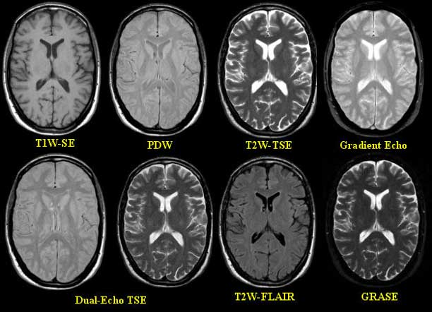
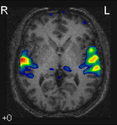
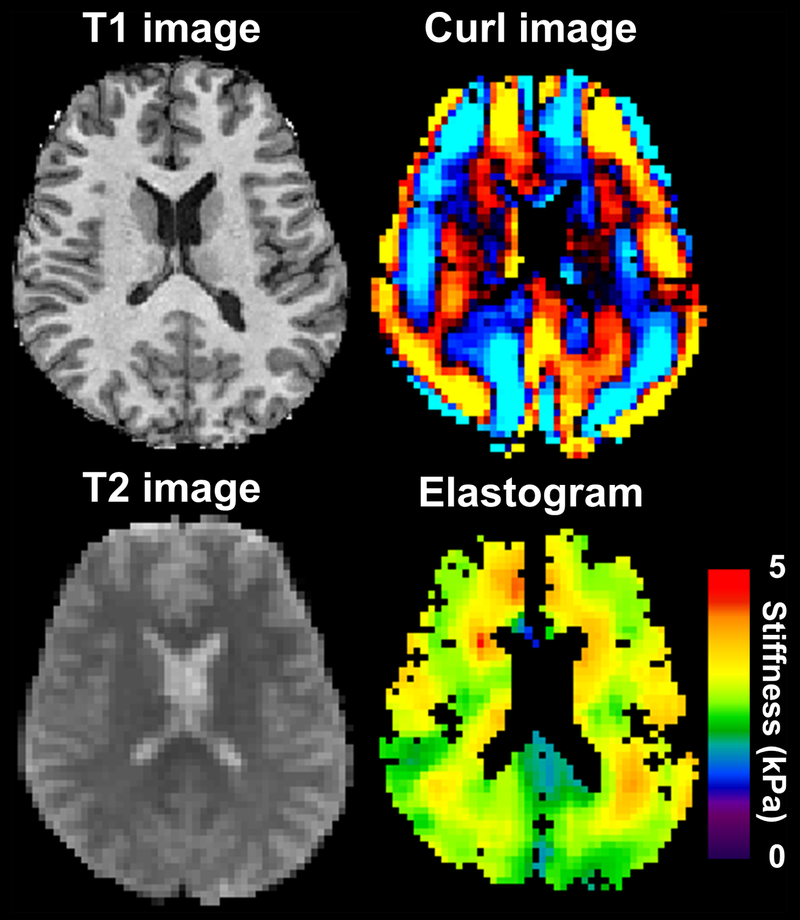
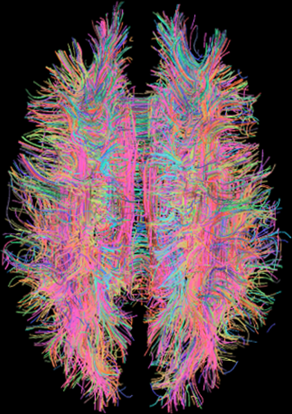
MRI Sequences of the Brain – Axial projectionFunctional MRIA T1 weighted image is shown in the top-left, and the corresponding T2 weighted magnitude image from the MRE data is shown in the bottom-left. The wave image is shown in the top-right panel, and the resulting elastogram is in the bottom-right panel. Author Murphy, Matthew et al.MRI diffusion tensor imaging of white matter tracts Courtesy Xavier Gigandet et. al. Wikimedia