
The Common Vein Copyright 2010
Definition
The septum pellucidum is a thin traingular membrane that partitions the frontal horn of the lateral ventricles. It runs vertically between the corpus callosum superiorly and the rostrum of the corpus callosum and the fornix inferiorly.
Structurally it consists of two layers that contain gray and white matter which are usually fused, but may be separated into two as a normal variant. This condition is called cavum septum pellucidum.
Functionally it participated in emotions of pleasure and rage, mood and sexual gratification.
Disease in the septum pellucidum results in cognitive impairment, as well as vision and coordination problems.
Diagnosis is by MRI and treatment is supportive.
 Septum Pellucidum Septum Pellucidum
Part of the Inverted C Rings – Navy Blue |
|
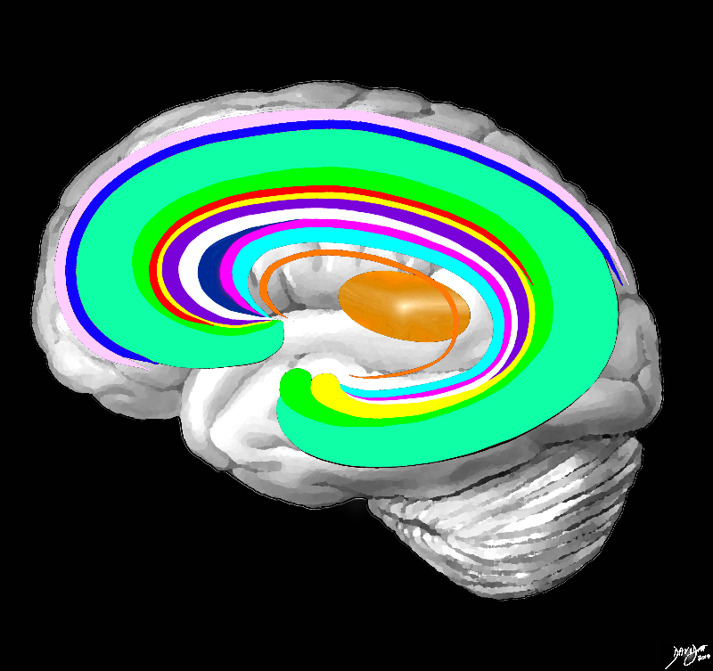
The forebrain has most of its components aligned in a series of inverted c- shaped rings starting from the outer membranes that culminate in the falx (pink), then extending inward smaller inner rings with each intimately connected to the others. The thalamus (dull orange) appears diagrammatically as the centre of these rings as seen from the sagittal view The outer ring is the falx (pink) followed by the sagittal sinus (blue) cerebral cortex (light green), cingulate gyrus (bright green) superiorly which becomes the parahippocampal gyrus inferiorly. The red ring represents the distribution of the main portion of the anterior cerebral artery. Next is the yellow ring which is the supracollosal gyrus (indusium griseum) superiorly and the hippocampus inferiorly. This is followed by the corpus callosum (purple) which enables the white ring of white matter to connect between hemispheres. The next ring is the thin bright pink ring which represents the fornix superiorly and the fimbria inferiorly. The innermost ring (light blue) represents the lateral horns of the ventricular system. The basal ganglia run just lateral to the lateral ventricles. The navy blue arrow headed structure is the septum pellucidum. Courtesy Ashley Davidoff copyright 2010 all rights reserved 83027b04g03.8s |
 Septum Pellucidum in between the Frontal Horns Septum Pellucidum in between the Frontal Horns |
|
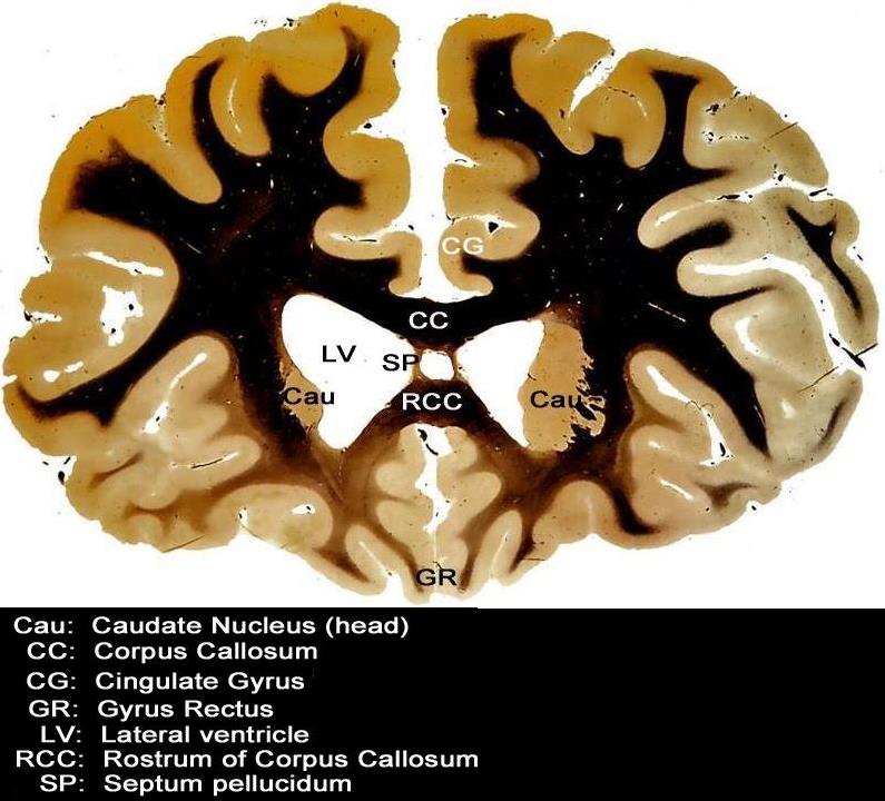
This coronal section through the forebrain is an anterior cut through the frontal lobes recognized by absence of the temporal horns, the presence of the rostrum of the corpus callosum (RCC), the cingulate gyrus (CG) superiorly and the gyrus rectus (GR) inferiorly. The visualized cortex is therefore part of the frontal lobe, and the ventricles (LV) that are visualized represent the frontal horns. The caudate nuclii (CN) are seen inferolateral to the frontal horns, and the septum pellucidum is seen medially Courtesy Department of Anatomy and Neurobiology at Boston University School of Medicine Dr. Jennifer Luebke, and Dr. Douglas Rosene 97342.C3.8L01 |
 Gross Anatomy of the Septum Pellucidum Gross Anatomy of the Septum Pellucidum |
|
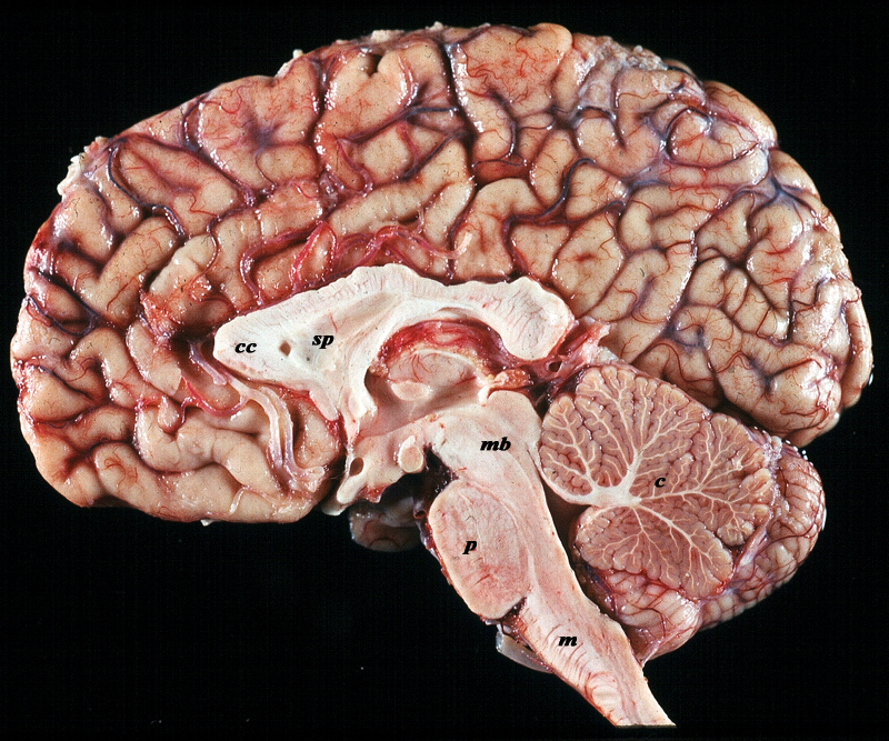
The midsagittal section view of brain reveals the distinctive shape position and character of the midline structures of the brain. The distinction between the character of the cerebral cortex which has a creamy color and the white matter exemplified by the corpus callosum (c) and septum pellucidum (sp) which are white, and the midbrain (mb) pons (p) and medulla (m) which are off white as opposed to the color of the cerebellum (c) which is light salmon pink is well demonstrated. The relative sizes of the forebrain, midbrain and hindbrain and their components are well appreciated in this section. Image Courtesy of Thomas W.Smith, MD; Department of Pathology, University of Massachusetts Medical School. 97805b02 |
 Septum Pellucidum in Coronal Projection Septum Pellucidum in Coronal Projection |
|
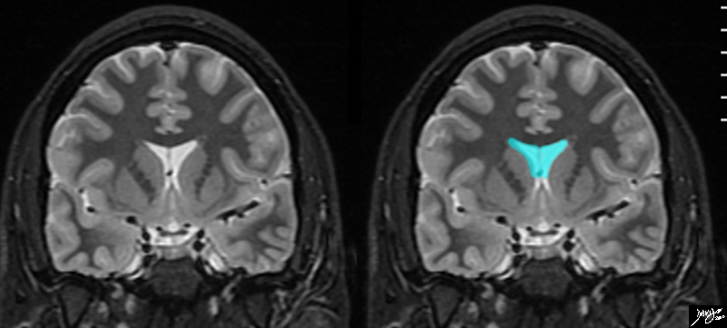
The frontal horns are seen in this coronal MRI using a STIR sequence. They are easily recognized as a T shaped midline structure, divided in the middle by the gracile septum pellucidum. The frontal horns course through the frontal lobes and above the anterior portion of the temporal lobes. At this level many of the components of the basal ganglia can be evaluated below and lateral to the frontal horns c Courtesy Ashley Davidoff MD copyright 2010 all rights reserved 89732c01.8s |
|
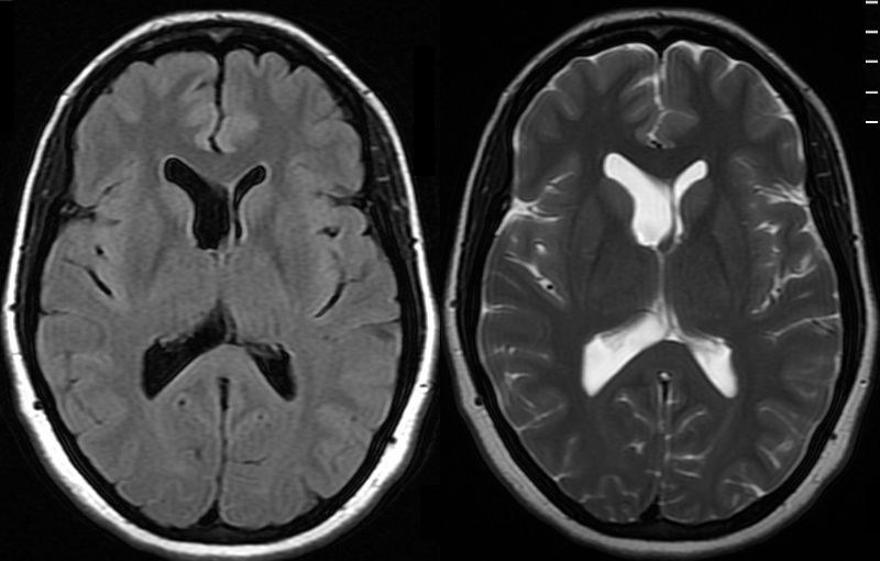
Asymmetry of the Lateral Ventricles – Minor Shift of the Septum Pellucidum – Normal Variant |
|
The MRI shows a FLAIR and a T2 weighted image revealing normal asymmetric development of the lateral ventricles in a 41 year old female. Courtesy Ashley Davidoff MD Copyright 2010 89044c |
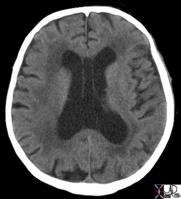 Cavum Septum Pellucidum Cavum Septum Pellucidum |
|
This CT demonstrates widening of the 2 septal laminae of the septum pellucidum in the lateral ventricles This condition is called cavum septum pellucidum which is a structural deformity of the septum pellucidum where there is a separation between its two leaflets (septal laminae) which have failed to fuse in the first 3-6 months of life. This condition is seen in 100% of fetuses , and about 85% fuse by 3-6 months Courtesy Ashley Davidoff MD Copyright All rights Reserved 49030 |
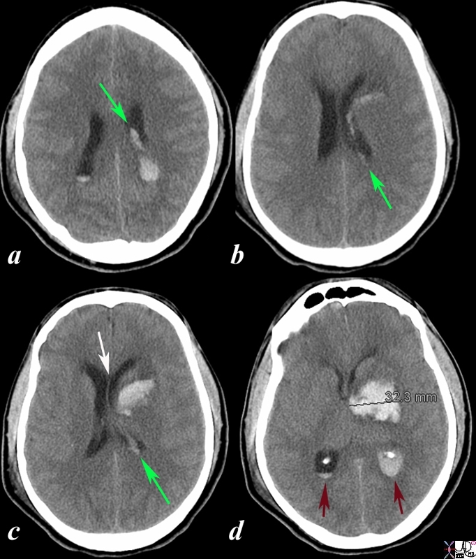 Acute Hemorrhagic Stroke of the Basal Ganglia Acute Hemorrhagic Stroke of the Basal Ganglia
Deviation of Septum Pellucidum |
|
The CT is from a 33year old male with an acute left basal ganglial hemorrhagic stroke. The CT shows a hyperdense accumulation of hemorrhage(d) complicated by extension or rupture of the hemorrhage into the ipsilateral choroid plexus (green arrows a,b,c) and hemorrhage into the ventricles with blood lying dependently in the occipital horns (maroon arrows in c) and midline shift with septum pellucidum (white arrow of the eyes (lenses overlaid in d) mass effect on the left frontal horn (d) and midline shift exemplified by the shift of the septum pellucidum (white arrow c). Image Courtesy Ashley Davidoff MD Copyright 2010 98551cL.8s |
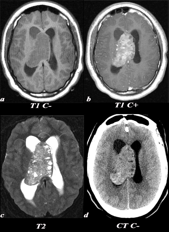 Central Neurocytoma Central Neurocytoma
Extends via the Lateral Ventricle to the Septum Pellucidum |
|
A 21 year old female was found to have papilledema on physical exam noted by her optometrist. MRI: T1 pre (T1 C- a) and post (T1 C+ b): Post contrast images demonstrate the heterogeneous enhancing nature of this mass. These images demonstrate centered in the right lateral ventricle with involvement of the septum pellucidum and partial extension into the left lateral ventricle. T2: T2 weighted images demonstrate internal areas of high signal consistent with cystic components. Also note the high T2 signal adjacent to the enlarged lateral ventricles which is a finding consistent with hydrocephalus, classically called transependymal flow of CSF. This unenhanced CT scan demonstrates also demonstrates the mass in the right lateral ventricle with similar features of heterogeneity and involvement of the septum pellucidum and partial extension into the left lateral ventricle. Notice the internal low density or cystic components and the higher density calcifications seen posteriorly. The lateral ventricles are enlarged consistent with resultant hydrocephalus. The diagnosis is consistent with a central neurocytoma. Image Courtesy Elisa Flower MD and AsimMian MD 97634c01.8s |