
Transverse or Axial View Concepts
The Common Vein Copyright 2010
Introduction
We continue with the theme of attempting to provide some reference points that are relatively easy to remember and provide an anchoring concepts for understanding further complexity.
In the axial or transverse view the ventricles provide an important reference point whether one is the in the fore brain, mid brain or hind brain territory, so we will familiarize the reader with the ventricular system in the transverse plain.
|
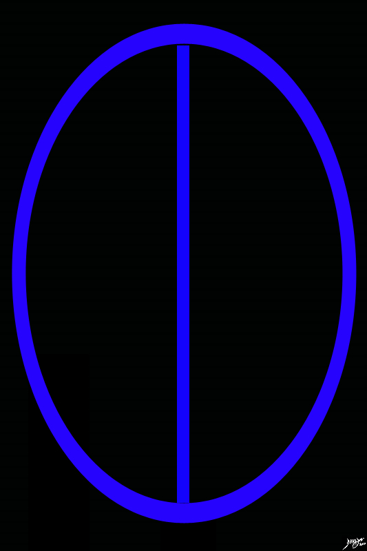
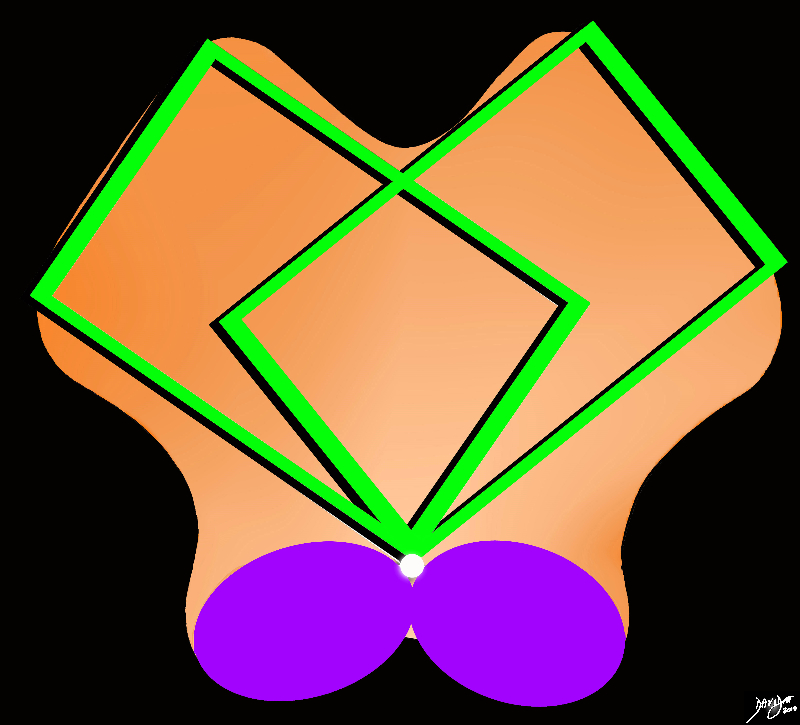
Axial Projection Superior projection The Two Cerebral hemispheres Divided by the Interhemispheric Fissure |
|
The diagram reflects the interhemispheric fissure as depicted in the axial plane dividing the brain into two halves. At the surface of the brain it is a division of the forebrain into the two cerbral hemisphres. By convention anterior is on the top and posterior on the bottom, while the patients right is to our left and vice versa for the patients left brain stick diagram Davidoff Art Copyright 2010 Courtesy Ash;ley Davidoff MD 93914.82s |
|
Backward C’s of the Forebrain Structures |
|
The initial approach to the axial images as we proceed deeper into the brain, and particulalrly the forebrain in this instance, is a series of back to front bilateral c’s, the shape of which is set by the lateral ventricles Davidoff art Courtesy Ashley Davidoff MD copyright 2010 93914.3ka09.8sd01 |
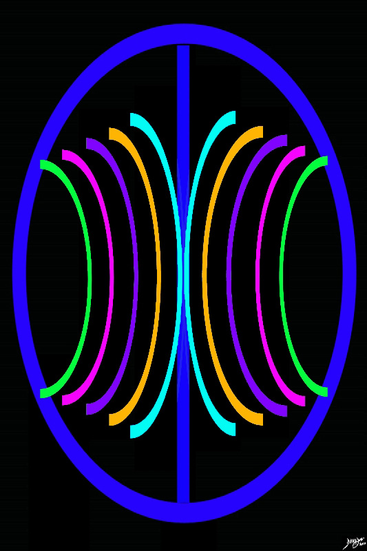
Knowledge of the structure of the ventricular system is crucial to provide a reference point for many of the internal structures of the brain
|
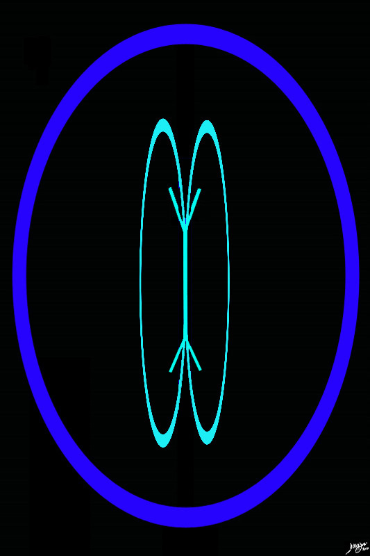
The Cranialmost Reference of the CSF system |
|
The axial system allows us to follow the CSF system and conceptually this provides a framework from which we can launch into the study of the anatomy Most superiorly the lateral ventricles first surface. They present as two relatively large almost ovoid structures on either side of the midline Davidoff art Courtesy Ashley Davidoff copyright 2010 93914.3kd03b02b01.81sd02 |
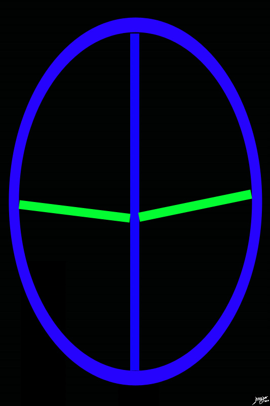
The sulci, fissures and gyri are numerable and complex but it is important to get to know some key border forming sulci. The central sulcus for example divides the fromntal lobe from the parietal lobe.
|
The Central Sulcus Separates the Frontal Lobes from the Parietal Lobes |
|
The diagram reflects the interhemispheric fissure as depicted in the axial plane dividing the brain into two halves. (blue). The sulci that separate the frontal lobes and the parietal lobes are called the central sulci (represented in green). Upward or forward of the central sulcus is the frontal lobe and backward or posterior are the parietal lobes. The occipital lobes and temporal lobes, midbrain and hindbrain are all mor caudal to this plane Davidoff art Courtesy Ashley Davidoff MD copyright 2010 93914.83s |
The internal structures of the brain are also complex but they seem to take on an abstract layering . This concept allows a framing of the structures and while nor an exact organization – it is a helpful way to conceptualise the brain.
For the midbrain pons and medulla, the shapes are very similar in the axial plane and subtle shapes and positioning and size of the components of the CSF system need to be visualised
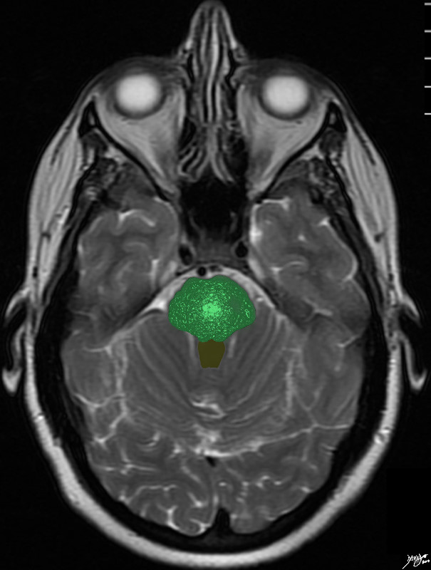
Mid Brain
Shape of the Midbrain |
|
The midbrain consists conceptually of twoanteroleaterally positioned rectancles, and two posterocentrally placed ovals (purple), surrounding the small aqueduct of Sylvius (white). Often it is the hint of the aqueduct relatively deep in the posterior wall of a rectangular structure that will help define the structure as the midbrain. Courtesy Ashley Davidoff MD copyright 2010 all rights reserved 94074b09b01a08.8s |
|
Peduncles Substantia Nigra Tegmentum Colliculi and Tectum |
|
The anterior border iof the midbrain incorporates the cerbral peduncles(green), and the substantia nigra (black – just posterior to the peduncles. Between the substantia nigra and the aqueduct is an area of the midbrain called the tegmentum (floor of the midbrain) The posterior end of the midbrain is bordered by the colliculi (purple) in the tectum (roof) of the nidbrain. Davidoff art Courtesy Ashley Davidoff MD copyright 2010 all rights reserved 94074b09b05b.83s |
The Hindbrain
The midbrain pons and medulla are diffficult to distinguish in the axial plane
Pons
The Pons |
|
The pons is difficult to recognise by intrinsic characteristics, but if you paint it green – render its matrix and paint the 4th ventricle brown it looks like a little bush in a flower pot – non descript in the axial plane – yes! Courtesy Ashley Davidoff copyright 2010 all rights reserved 94084.3kb08.81s |
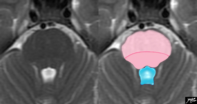
Characteristic 4th Ventricle |
|
TheT2 weighted MRI is a magnified view of the pons recognized commonly in the axial projection by its relationship to the 4th ventricle posteriorly which has a characteristic shape looking like a puffed up crown (blue) The pons consists of an anterior portion called the ventral pons (light pink) and the dorsal portion called the tegmentum ((darker pink). The line of distinction is vague and the dark pink line is drawn in for clarity. Courtesy Ashley Davidoff MD copyright 2010 all rights reserved 94084.3kdc.82sd01 |
Medulla
|
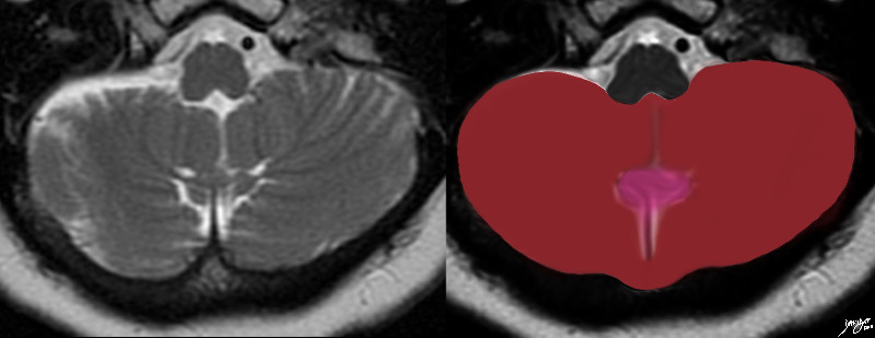
The Medulla 4 Ice Cream Cones in a Row |
|
The medulla at its classic location has some subtle shape diferences when compared to the midbrain and pons mostly accentuated by the olives (green)which are the second set of ice cream shaped wedges . The medial set of ice cream cones that are light pink are the medullary pyramids Posteriorly – the salmon pink bilobed section is called the tegmentum Courtesy Ashley DAvidoff MD copyright 2010 all rights reserved 94086b01.3kb02bb04.8s |
The Cerebellum
The cerebellum is an easily recognized as posterior fossa structure that looks like an upseide down spinning top
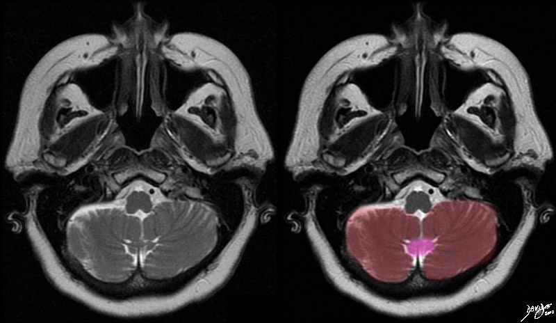
A Heart or a Top |
|
The transverse Tt2 weighted MRI through the main portion of the cerebellum in the posterior cranial fossa reflects a heart shaped structure created with some artistic licence. The cerebellum consists of two cerebellar hemispheres and a central worm like vermis Courtesy Ashley Davidoff MD copyright 2010 all rights reserved 49037.3kb01c.8s |
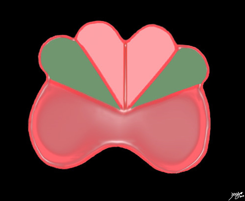
The Hemispheres and the Vermis |
|
The T2 weighted MRI shows the 2 major components of the cerebellum – the central vermis (pink), and the two cerebellar hemispheres. The cerebellum consists of two cerebellar hemispheres and a central worm like vermis Courtesy Ashley Davidoff MD copyright 2010 all rights reserved 49037c.8s |