Internal Carotid Artery
The Common Vein Copyright 2010
Definition
The internal carotid artery is part of one of the two main systems that supply the brain with blood.
It orginates as a branch of the common carotid artery at the the level of the upper border of the thyroid cartilage.
The internal carotid artery arises from the carotid bifurcation at the level of vertebra C4, running through the anterior edge of the sternocleidomastoid. In the maxillo-pharyngeal space it ascends anteriorly and internally to the internal jugular vein. This is the C1 segment.
It proceeds cranially anterior to the transverse processes of three cervical vertebra to the inferior surface of the petrous portion of the temporal bone, after which it enters the carotid canal.
It accesses the base of the skull through the carotid canal, anteriorly to the jugular foramen – C2 segment.
Thereafter it curves anteriorly, and and maintains a horizontal course travelling slightly medially within the carotid canal. It proceeds cranially and crosses the upper part of the foramen lacerum, entering the the middle cranial fossa.
Thus, it will enter the petrous portion of the temporal bone while in the carotid canal, being accompanied by a sympathetic plexus from the cervical chain.
This segment has a:
Vertical/Ascending segment anteriorly and internally to the tympanic cavity.
Genu segment in the axis of the petrous portion (curvature)
Horizontal segment that enters the cavernous sinus above the foramen lacerum
The C3 segment is a short segment that begins above the foramen lacerum, ending at the petrolingual ligament.
From here and until it perforates the dura – the C4 segment – describes a siphon and enters the cavernous sinus, and crosses laterally the posterior and anterior clinoid processes.In this segment, the artery is surrounded by nerves of the sympathetic trunk and the abducent nerve
It thus becomes intradural, until it enters the subarachnoid space – this short segment is called the C5 segment. As it gets into the subarachnoid space – C6 segment – it gives its main collateral – the ophthalmic artery.
Finally, C7 segment is when it passes between nerves II and III and the anterior perforated substance. It provides four terminal branches – the cerebral arteries:
anterior cerebral artery
middle cerebral artery
anterior choroidal artery
posterior communicating artery
Origins of the Internal Carotid Artery |
|
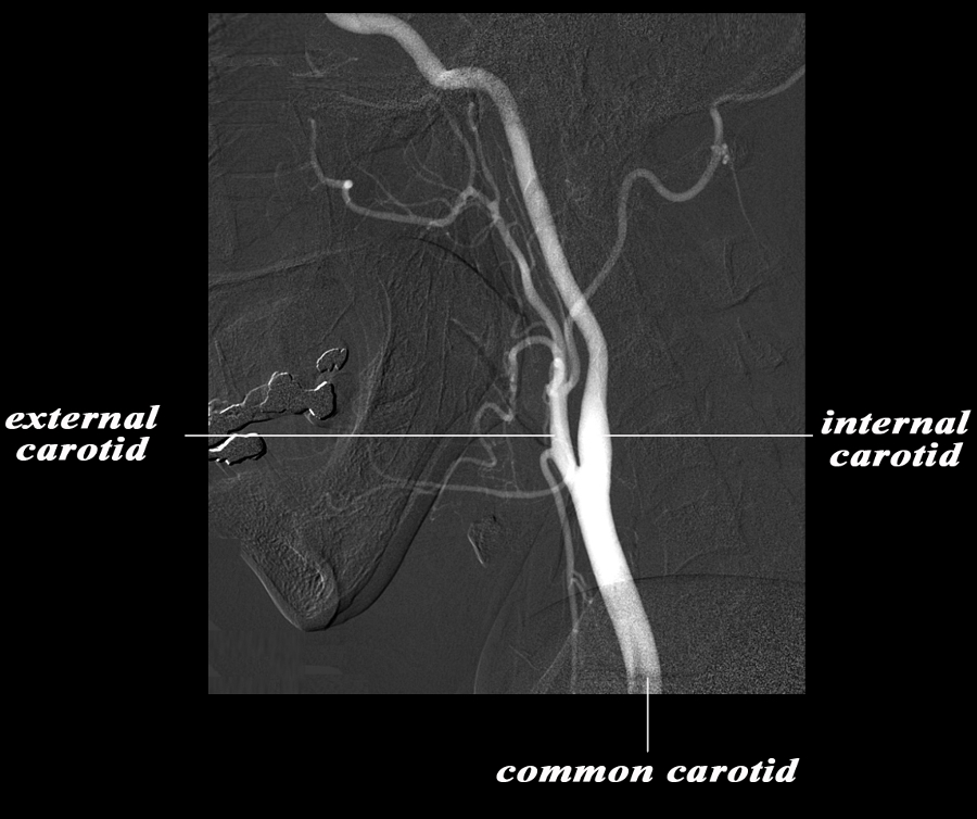
The angiogram demonstrates the carotid system in the lateral projection. The common carotid artery bifurcates in the base of the neck into the external and internal carotid artery. Basically the internal carotid artery supplies the brain and the external supplies the structures of the face and muscles and skin of the cranium. The carotid system supplies the brain from the internal carotid – a branch of the common carotid which arises from the aorta. Courtesy Ram Chavalli MD 49400bc01.8s |
The segments of the internal carotid artery inlude;
cervical segment
petrous segment – extends from the base of the skull to the apex of the petrous bone and enters the cranial vault via the foramen lacerum
cavernous segment – passes through the cavernous sinus
supraclinoid segment after the cavernous segment perforates the dura, it becomes the supraclinoisd segment which ends shortly when it bifurcates into the middle and anterior cerebral arteries.
|
Segments of the Internal Carotid Artery |
|
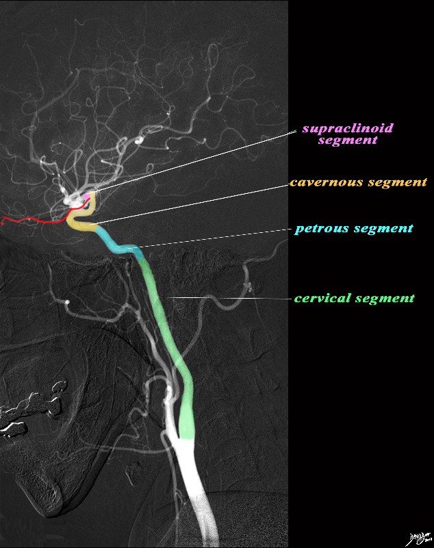
The angiogram demonstrates the carotid system in the lateral projection. The common carotid artery bifurcates in the base of the neck into the external and internal carotid artery. Basically the internal carotid artery supplies the brain and the external supplies the structures of the face and muscles and skin of the cranium. The internal carotid artery is divided into segments. The cervical segment (green) extends from the bifurcation to the base of the skull. The petrous segment (teal blue extends from the base of the skull to the apex of the petrous bone and enters the cranial vault via the foramen lacerum. The cavernous segment (light orange) passes through the cavernous sinus and exits by penetrating the dura. The ophthalmic artery (red) originates at the junction of the cavernous and supraclinoid segment. The supraclinoid segment materializes when the cavernous portion leave the dura. After a short distance it terminates by bifurcating into the middle and anterior cerebral arteries. code brain anatomy neuroanatomy normal blood supply common carotid internal carotid external carotid internal carotid artery segments cervical segment petrous segment cavernous segment supraclinoid segment middle cerebral artery anterior cerebral artery angiogram angiography selective Image courtesy Ram Chavalli MD Artistic rendering Davidoff MD Copyright 2010 49400bg.8s |
Angiogram in the Frontal Projection |
|
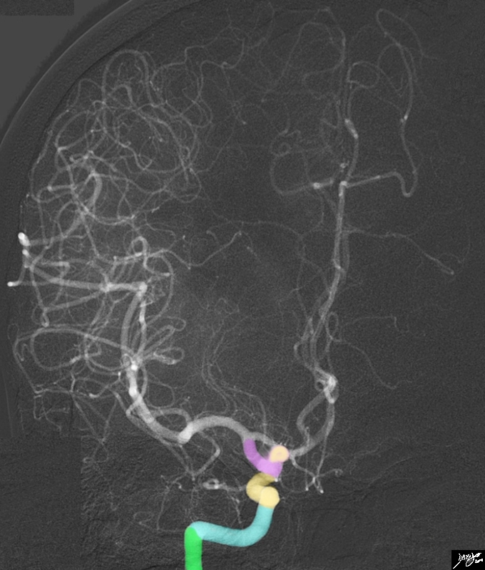
The intracranial angiogram in A-P projection demonstrates the segments of the internal carotid system The terminal end of the cervical segment (green) extends from the bifurcation to the base of the skull. The petrous segment (teal blue) extends from the base of the skull to the apex of the petrous bone and enters the cranial vault via the foramen lacerum. The cavernous segment (light orange) passes through the cavernous sinus and exits by penetrating the dura. The supraclinoid segment materializes when the cavernous portion leave the dura. After a short distance it terminates by bifurcating into the middle and anterior cerebral arteries. Image courtesy Ram Chavalli MD Artistic rendering Davidoff MD Copyright 2010 49412b02.8s |
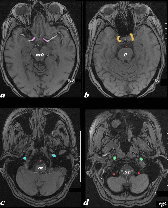
Segments of the Internal Carotid Axial Projection |
|
The T1 weighted MRI following contrast in axial projection shows the segments of the internal carotid artery. Starting from image (a) the most cranial cut, the distal tip of supraclinoid segment (pink) is recognized by the division into the middle cerebral arteries (white). The anterior cerebral arteries are nor seen in this section. The supraclinoid segment materializes when the cavernous portion leave the dura. After a short distance it terminates by bifurcating into the middle and anterior cerebral arteries. At this level the midbrain (mb) is visible. The cavernous segment (light orange) is seen in (b). It passes through the cavernous sinus and exits by penetrating the dura. At this level the pons (p) and basilar artery (red) are visible. The petrous segment (teal blue) extends from the base of the skull to the apex of the petrous bone and enters the cranial vault via the foramen lacerum. It is seen in (c). At this level part of the medulla (m) and basilar artery (red) are visible. The cervical segment (green) extends from the bifurcation to the base of the skull and is seen in (d). At this level the spinal cord and the two and vertebral arteries (red) are visible. Courtesy Ashley Davidoff MD Copyright 2010 49264C03.8s |
|
Intracranial Branches |
|
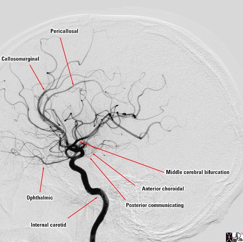
The selective of the internal carotid artery in the lateral projection reveals the major intracranial branches this vessel; The ophthalmic artery is is the first branch of the internal carotid artery distal to the cavernous sinus and it runs anteriorly to supply the eye, nose and meninges The ICA then gives off the posterior communicating artery that joins the circle of Willis followed by the anterior choroidal artery that supplies the choroid plexus but also many other forebrain structures including the optic chiasm, internal capsule, globus pallidus, tail of the caudate nucleus , amygdala. It also supplies midbrain structures including substantia nigra, red nucleus and crus cerebri After this it divides into a anterior cerebral artery that supplies the anterior and medial portion of the frontal lobes . It divides into the callosomarginal artery pericallosal artery middle cerebral artery that supplies the frontal region laterally, the parietal lobe, and the temporal lobe Courtesy Elisa Flower MD and Alex Norbash MD copyright 2010 97626b01.8s |
|
Intracranial Portion of the ICA – A-P Projection |
|
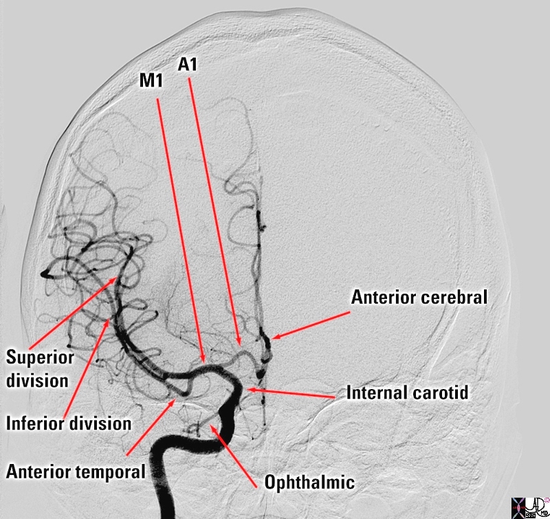
The selective of the internal carotid artery in the anteroposterior projection shows the major intracranial branches; The ophthalmic artery is is the first branch of the internal carotid artery distal to the cavernous sinus and it runs anteriorly to supply the eye, nose and meninges The ICA divides into the anterior cerebral artery (ACA) and the first segment is called the A1 segment that supplies the anterior and medial portion of the frontal lobes . middle cerebral artery (MCA) that supplies the frontal region laterally, the parietal lobe, and the temporal lobe Its first portion is called the M1 segment and it divides into two basic segments Superior division Inferior division The other vessel that can be seen in this view is the anterior temporal artery Courtesy Elisa Flower MD and Alex Norbash MD Copyright 2010 97628b.8s |
Initially in the cranial cavity, the inernal carotid artery is between layers of the dura mater, thus forming and being an intimate part of the cavernous sinus.
The artery then ascends toward the the posterior clinoid process, makes an s-shape, heads into the portion of the dura mater that forms the roof of the cavernous sinus, on the medial edge of the anterior clinoids.
It courses between the optic nerve and the oculomotor nerve reaching the anterior perforated substance. At this point it divides into its terminal branches – the middle and anterior cerebral arteries.
Applied Biology – Manifestations of Disease in the Internal Carotid
|
High Grade Stenosis of the Intenal Carotid Artery |
|
The selective angiogram of the the common carotid shows a high grade subtotal occlusion of the origin of the internal carotid artery. This is likely due to atherosclerosis. 10218 Courtesy Ashley Davidoff |

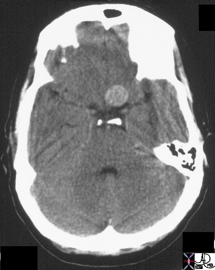
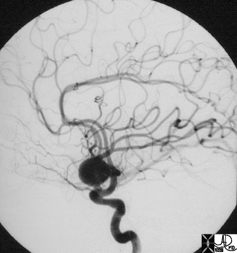
Aneurysm of Supraclinoid Portion of the ICA |
|
The selective right common carotid angiogram shows a small and broad based internal carotid artery aneurysm (purple) arising form the supraclinoid portion (salmon pink). The ophthalmic artery (red) that arises from the border between the supraclinoid portion and the cavernous portion of the internal carotid artery is noted Other components noted include the cavernous portion (orange), the division into the anterior cerebral arteries, (bright green) and middle cerebral artery (dark green). Image Courtesy Ram Chavalli MD Copyright 2010 All rights reserved 49409Sc01.8 |
Aneurysm |
| The CT on the right shows an enhancing mass near the pituitary fossa confirmed to be an aneurysm of the anterior communicating artery on angiography.
20258 Courtesy Ashley Davidoff MD |
Complete Circulatory Failure
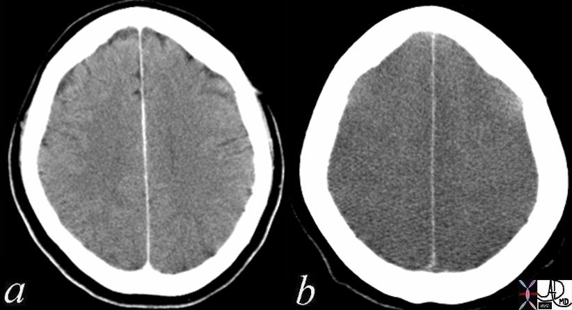
Normal – 4 months Prior and Folllowing Cardiopulmonary Arrest Loss of Gray White Differentiation |
|
The CT is from a 52 year old female who had a CT 4 months prior tto a cardiac arrest (a) showing subtle but definite difference in the attenuation difference between gray and white matter. After the cardiac arrest, she suffered anoxic injury to the brain resulting in extensive and global swelling of the brain and both gray and white matter have the same attenuation. The distinction between the two cannot be made and this finding is consistent with global anoxic injury. After 10-15 seconds of anoxia from cardiac arrest, loss of conciousness occurs and after 5 minutes of arrest, irreversible brain injury occurs. The gray matter of the brain, particularly the frontal lobes have highest metabolic needs. The occipital, parietal, and temporal lobes and basal ganglia and cerebellum have lesser needs. The brainstem has the lowest oxygen needs Courtesy Ashley Davidoff MD 70134c01 |
|
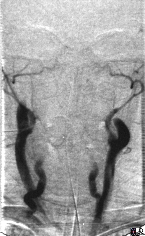
Total Constriction of the Internal Carotid Arteries |
| This image represents the sad ending of life for this individual. The extracranial vessels are all occluded and there is no intarcerebral flow or perfusion. These findings are compatible with brain death.
Courteys Ashley Davidoff MD Copyright 2010 22772 |