
Copyright 2010
Definition
The choroid plexus is a structure found in all four ventricles structurally characterized by its delicate yet irregular appearance and their common occurrence of calcification particulalrly ithe choroid plexus in the atria.. At a histological level it consists of a rich network of capillaries surrounded by an epithelieal layer whichis responsible for the production of the CSF
Functionally it is responsible for the production of cerespinal fluid. Its name is quite ambivalent as it derives from the Latin word chorion which means delicate and plexus which means a knot.
Diseases that affect the choroid plexus include choroid plexus cysts which are quite commonly seen in utero as an isolated finding but smetimes also seen chromlsomal abnormalities. These often resorb in the latter part of the pregnancy. Choroid plexus tumors are rare
 Choroid Plexus 20X Choroid Plexus 20X |
|
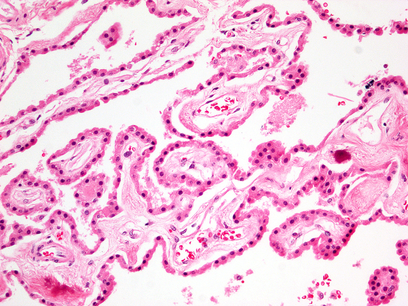
This H&E histological section at 20X magnification reveals normal choroid plexus characterized by folds or villi consisting of simple cuboidal epithelium surrounding networks of capillaries Image Courtesy of Thomas W.Smith, MD; Department of Pathology, University of Massachusetts Medical School. 97383 |
 Choroid Plexus 60X Choroid Plexus 60X |
|
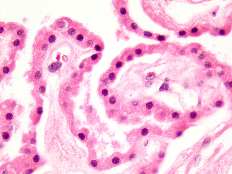
This H&E histological section at 60X magnification reveals normal choroid plexus characterized by folds or villi consisting of simple cuboidal epithelium surrounding networks of capillaries. Some of the secretion with exfoliation is seen within one of the villi. Image Courtesy of Thomas W.Smith, MD; Department of Pathology, University of Massachusetts Medical School. 97384 |
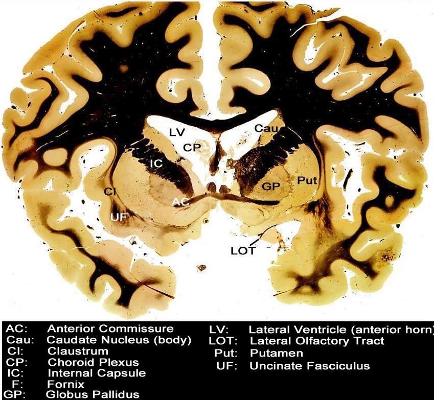
Choroid Plexus within the LAteral Ventricles |
| Courtesy Department of Anatomy and Neurobiology at Boston University School of Medicine Dr. Jennifer Luebke , and Dr. Douglas Rosene 97345.C6.8L01 |
 Normal Choroid Plexus – T2 weighted MRI Normal Choroid Plexus – T2 weighted MRI |
|
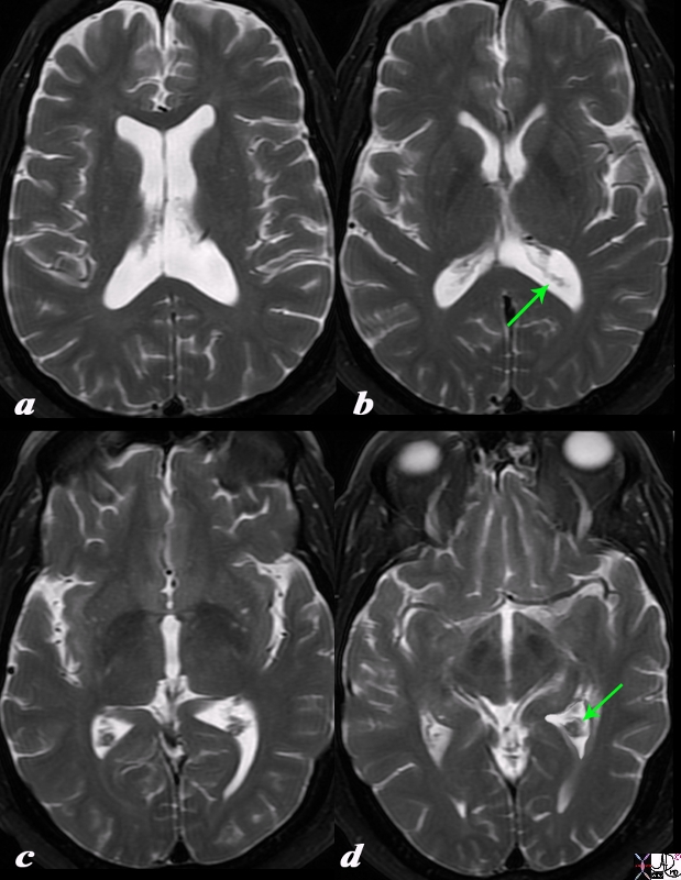
The T2 weighted MRI axial series is from a normal 69year old man it reveals the choroid pleaxus as fine strands and lobular tissue in the body of the ventricles (a) , in the occipital horns (b green arrow) in the atria (c) and extending into the temporal horns (green arrow). Courtesy Ashley Davidoff MD Copyright 2010 98204cL.8s |
|
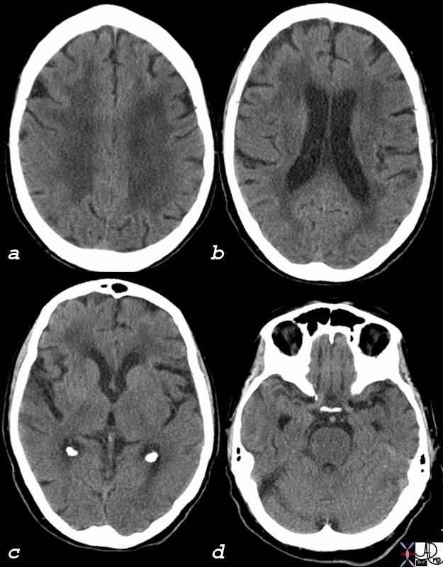
Age related Atrophy Temporal Horns become Visible (d) |
|
The CT scan of this 92 year old man reveals normal involutional change of the brain including perivntricular lucency(a)suggestive of microangiopathic change, mild dilatation of the ventricles (b) with deepening of the sulci and prominence of the gyri (abc) and ability to identify the temporal horns (d), all signs of brain atrophy. Note also the normal calcification of the choroid plexus in the atria (c) which is found very commonly even in young patients. Image Courtesy Ashley Davidoff MD 75932c01 |
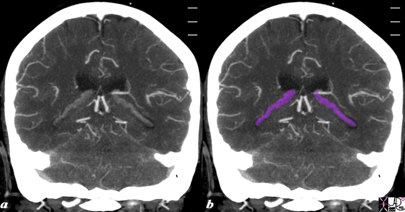 Choroid Plexus in the Temporal Lobe Choroid Plexus in the Temporal Lobe |
|
The reconstructed CTA scan in the coronal plane reveals the choroid plexus (purple) as fine knotty strands extending from the atrium to the temporal horns bilaterally. Image Courtesy Ashley Davidoff MD Copyright 2010 76532c.8s |
Applied Biology – Diseases
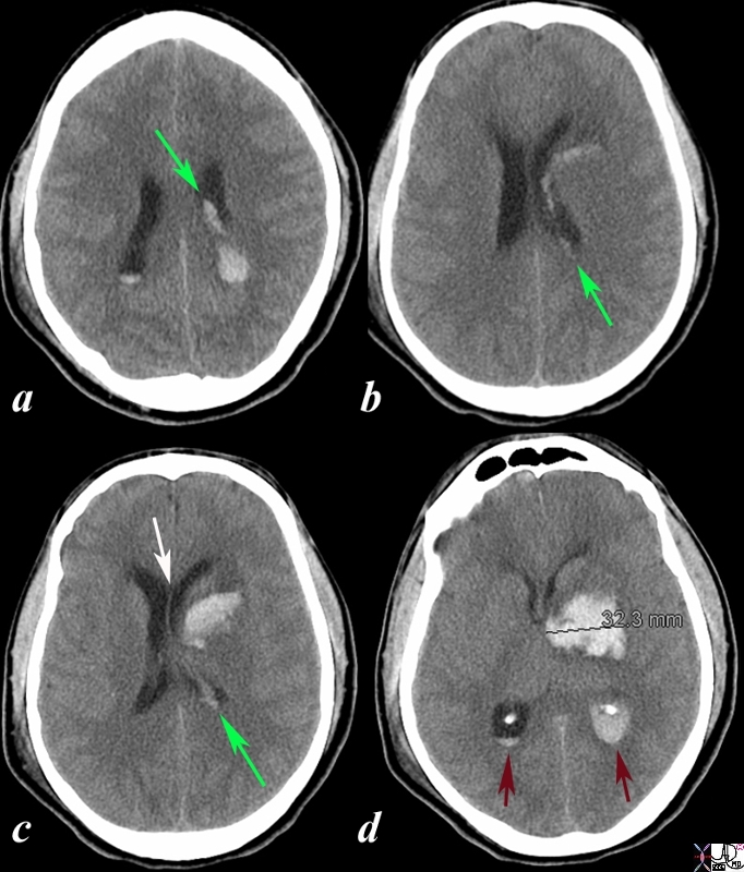 Acute Hemorrhagic Stroke of the BAsal Ganglia Acute Hemorrhagic Stroke of the BAsal Ganglia
Rupture into the Choroid Plexus |
|
The CT is from a 33year old male with an acute left basal ganglial hemorrhagic stroke. The CT shows a hyperdense accumulation of hemorrhage(d) complicated by extension or rupture of the hemorrhage into the ipsilateral choroid plexus (green arrows a,b,c) and hemorrhage into the ventricles with blood lying dependently in the occipital horns (maroon arrows in c) and midline shift with septum pellucidum (white arrow of the eyes (lenses overlaid in d) mass effect on the left frontal horn (d) and midline shift exemplified by the shift of the septum pellucidum (white arrow c). Image Courtesy Ashley Davidoff MD Copyright 2010 98551cL.8s |
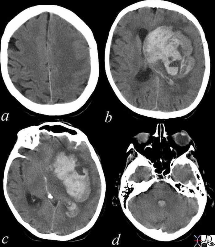 Acute Cerebral Hemorrhage with Mass Effect and Rupture into the Ventricles Acute Cerebral Hemorrhage with Mass Effect and Rupture into the Ventricles |
| This CT shows an acute hemorrhagic event originating in the frontoparietal region of the left cerebral hemisphere causing significant mass effect by compressing and displacing the ipsilateral lateral ventricle with significant midline shift.The hemorrhage has ruptured into the ipsilateral lateral ventricle and blood can be seen within the choroid plexus (b) in the posterior horn (c) as well as the 4th ventricle (d).
The ipsilateral edema has caused loss of the gray white matter interface in the left parietal lobe Courtesy Ashley Davidoff MD Copyright 2010 72143c01 |
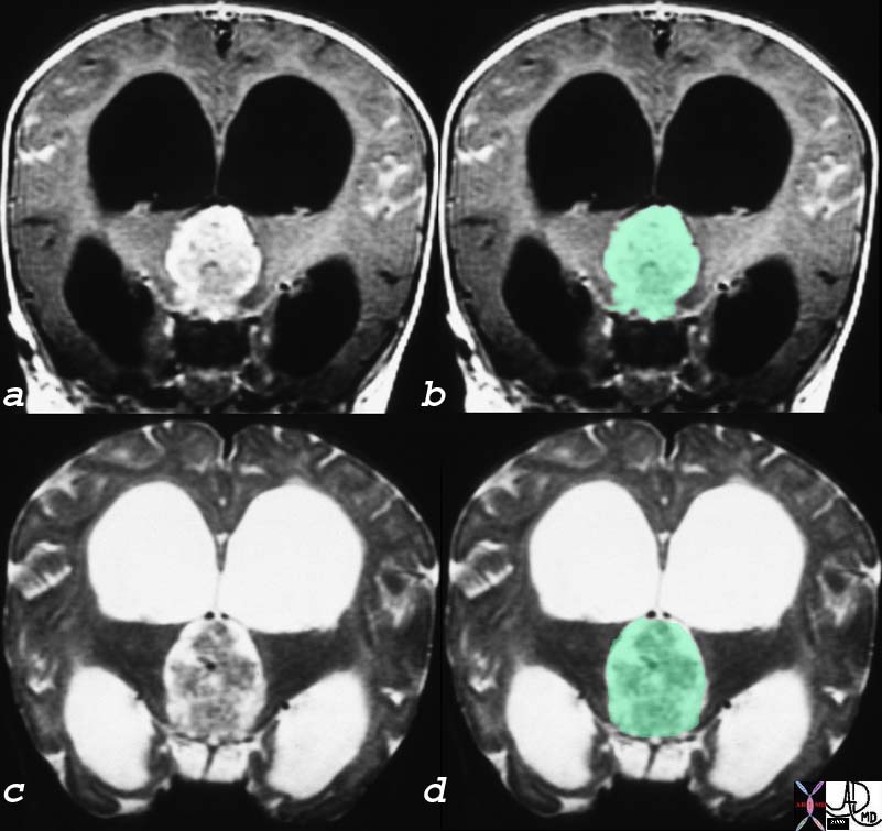
Papilloma of the Choroid Plexus Causing Hydrocephalus and Headache |
| The coronal MRI shows a gadolinium enhanced T1 weighted image (a,b) with black CSF in the dilated ventricles and a T2 weighted image with bright CSF (c,d). A large mass in the third ventricle (overlaid ingreen) dominates the study and is complicated by severe hydrocephalus. The mass was a choroid plexus papilloma of the third ventricle. In patients with brain tumor one of the causes of headache is hydrocephalus. This is an extreme case of hydrocephalus.23223c01 Courtesy James Donnelly MD |