
The Common Vein Copyright 2010
Definition
The forebrain lies as the topmost layer of the brain both structurally and functionally. In early development it lies as the frontward swelling, as one of the three swellings that develop from the evolving neural tube. It subsequently divides into two more components called the telencephalon and the diencephalon. The telencephalon evolves into the two cerebral hemispheres which contribute 80-90% of the brain substance while the diencephalon develops into the thalamus and hypothalamus. The thalamus acts as a relay station for the sensory input from our external and internal environment and sends the information to the cortex. The hypothalamus helps control and integrate autonomic activityand endocrine activity.
 Cerebrum or Telencephalon – Part of the Forebrain Cerebrum or Telencephalon – Part of the Forebrain
Crebral Hemispheres and Basal Ganglia |
|
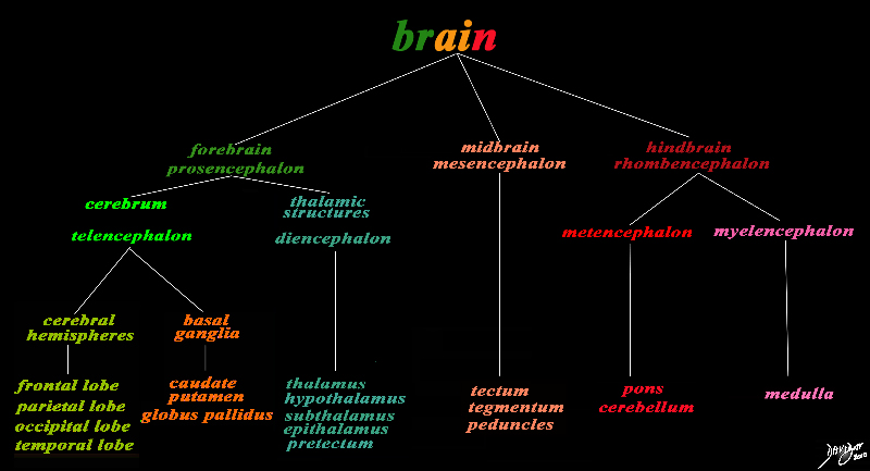
The basic and simplest classification of the brain into forebrain midbrain and hindbrain is shown in this diagram and advanced to a more complex tree using the embryological and evolutionary terminologies. The forebrain consists of the cerebrum also called the prosencephalon, which contains the more advanced form of the brain and the thalamic structures which contain more basic structures. The cerebrum (telencephalon) itself consists of two cerebral hemispheres and paired basal ganglial structures. Each cerebral hemisphere will have gray and white matter distributed in the frontal parietal temporal and occipital lobe, with the basal ganglia being part of the gray matter deep in the cerebral hemispheres. The most important thalamic structures arising from the diencephalons include the thalamus itself and the hypothalamus. The midbrain (mesencepaholon) consists of the tectum tegmentum and cerebral peduncles. The hindbrain has two major branch points based on the evolutionary development. The pons and cerebellum(part of the metencephalon) are grouped and the medulla (part of the myelencephalon is the second branch. Courtesy Ashley Davidoff MD Copyright 2010 All rights reserved 97686.8s |
 Cerebrum – Cerebral Hemisphere and Basal Ganglia Cerebrum – Cerebral Hemisphere and Basal Ganglia |
|
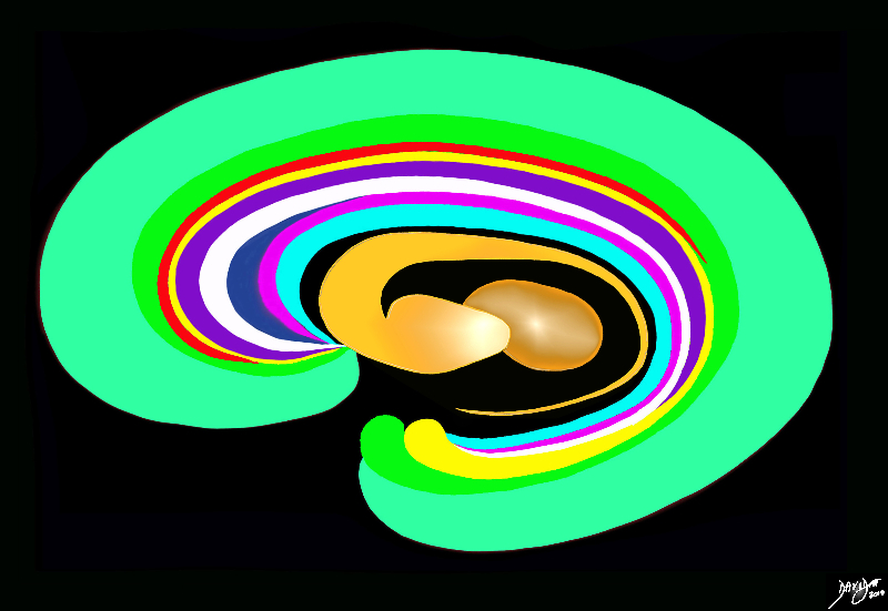
The orange c shaped ring lies lateral and to some extent inferior to the lateral ventricles typified by the light blue ring and consists of the thalamus and basal ganglia including the globus pallidus, putamen, head of the caudate nucleus, and tail of the caudate nucleus. This diagram infers the concept of recurring inverted C shaped structures that are layered both from deep to superficial but also to some extent medial to lateral. Courtesy Ashley Davidoff MD Copyright 2010 All rights reserved 93907d13b05b01.8s |
Cerebral Cortex
The cerebral cortex is 2-4 mm thick, composed of neurons and unmyelinated fibers. The unmyelinated fibers are what are commonly referred to as “grey matter”. White matter consists of the myelinated axons connecting nerve cells for signaling. Signals travel to and from gray matter through white matter.
The surface is composed of folds called sulci and gyri.
The majority (2/3) of the cerebral cortex is buried within these folds.
The cerebral cortex plays a role in memory, as well as executive cognitive functions and language. The cortex receives input exclusively from thalamic nuclei.
Diseases of the cerebral cortex include Alzheimer’s, Wilson’s disease, and multiple sclerosis. Diagnosis of the cerebral cortex involves observation of cognitive impairment and neuroimaging tests. Pharmacological treatment is used to mitigate symptoms but dysfunction of the cerebral cortex is degenerative.
 Cerebral Hemisphere – External View Cerebral Hemisphere – External View |
|
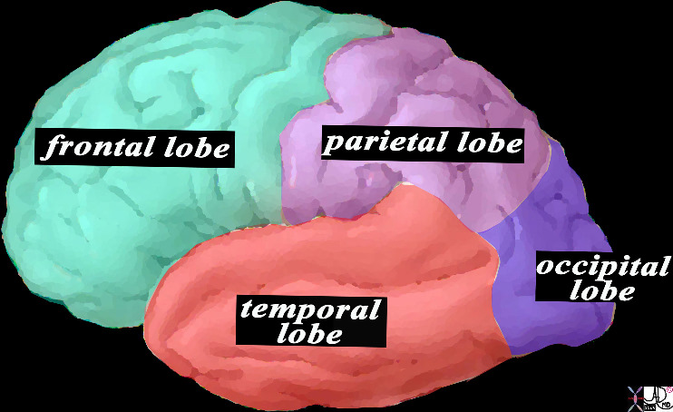
This is a diagram looking at part of the the forebrain from the side showing the anteriorly placed frontal lobe, inferiorly placed temporal lobe posteriorly placed occipital lobe and superiorly placed occipital lobe. In this view the size of the frontal lobe dominates and in fact it is the largest portion of the forebrain. Courtesy Ashley Davidoff MD copyright 2010 all rights reserved83029d13.8s |
Frontal Lobe
The frontal lobe is located at the front of the cerebral hemisphere and in front of the temporal and parietal lobes. The frontal lobe has been found to have a role in executive thought processes and decision-making behavior. Dysfunction in this area can lead to schizophrenia and severe depression as well as personality and emotional changes. Diagnosis involves completion of various general cognitive response tests as well as tests for specific cognitive deficits.
Parietal Lobe
It is above the occipital lobe but behind the frontal lobe and in between the temporal lobes. It is primarily composed of interneurons and integrates information from various receptors, primarily for the purpose of orienting in space and mapping the environment. Gerstmann’s syndrome, Balint’s syndrome, and hemispheral neglect are all associated with parietal lobe dysfunction. Treatment would involve therapy in less extreme cases, and permanent care assistance for those with extreme damage.
Temporal Lobe
The temporal lobe is located at the side of the brain beneath the Sylvian fissure. It is involved in auditory perception (hearing and speech) as well as memory. The hippocampus is located within the temporal lobe. Dysfunction in the temporal lobe leads to memory impairment as well as difficulties producing and understanding speech. Damage also causes extreme personality changes. Dysfunction is diagnosed through the observation of imapairment, cognitive and speech related tests, and neuroimaging to identify the cause. Treatment includes speech therapy as well as pharmocological agents to combat personality changes.
Occipital Lobe
The occipital lobe is at the rear of the skull, below the parietal lobe and it is the smallest of the four lobes. It is responsible for visual processing. Dysfunction can result in vision loss or hallucinations. Less severe dysfunction results in the inability to recognize or accurately perceive objects. Treatment is often surgical for the removal of lesions.
Basal Ganglia
The basal ganglia are a group of nuclei connecting structures within the forebrain and hindbrain. Two sets of ganglia are mirrored in the hemispheres of the brain. Dysfunction of the basal ganglia is linked to ADHD, cerebral palsy, OCD, and Parkinson’s. Treatment and diagnostic measures depend on the cause of disease. Treatment is often medicinal and diagnostic relies on MRI and other neuroimaging techniques.
 The Basal Ganglia The Basal Ganglia |
|
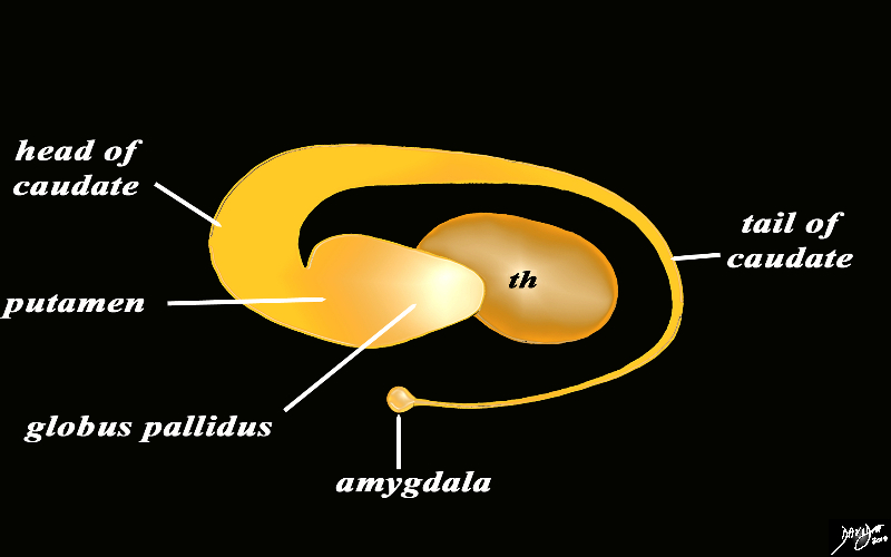
The basal ganglia lie both in the forebrain and in the midbrain. In the forebrain they are distributed in paraventricular fashion almost in an inverted C fashion. In the conceptual diagrams we have colored the inverted “C” that abuts the ventricular system orange, and this includes the thalamus and the basal ganglia. The basal ganglia that lie in the forebrain include the globus pallidus, putamen, head of the caudate nucleus, tail of the caudate nucleus and the amygdala. The amygdala appears to be part of the limbic system and the basal ganglia. Courtesy Ashley Davidoff MD Copyright 2010 All rights reserved 93907d13b05dL3.8s |