
The Common Vein Copyright 2010
Definition
The dural sinuses are vascular structures of the brain that particpate in venous drainage and CSF drainage of the brain, being structurally unique in the venous sytem since they are located and protected between two layers of dura. The major sinuses include the superior sagittal sinus, inferior sagittal sinus, straight sinus, sigmoid and transverse sinuses and the cavernous sinus.
The function of the dural sinuses is to provide venous drainage and CSF drainage for the brain. They receive blood from the cerebral veins, and CSF from the arachnoid granulations, and empty both products into the internal jugular vein for recirculation.
Diseases that affect the dural sinuses include trauma, subdural hematoma and venous sinus thrombosis.
Diagnosis of disease is usually by imaging the veins with contarast enhanced CT and MRI, or via the venous phases of arteriography.
Treatment depends on the cause of disease, but venous thrombosis is treated by anticoagulation while traumatic events treated by surgery with evacuation when hemorrhage limits function or raises the pressure to concerning levels.
Structural Principles
|
The Major Sinuses |
|
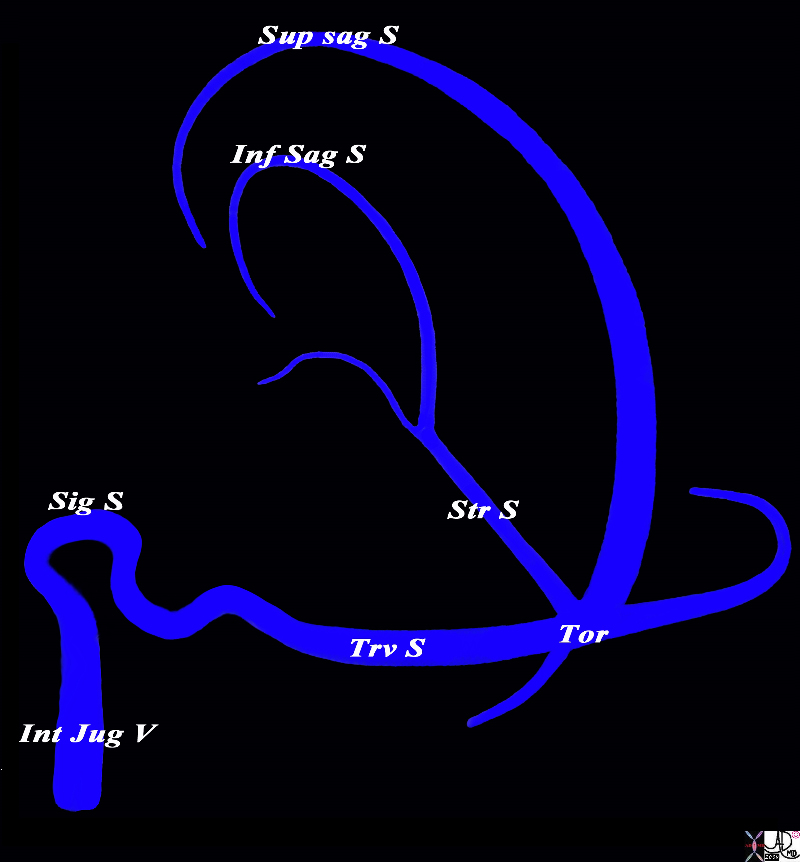
The diagram shows the basic plan of the dural sinuses and venous drainage of the brain in the parasagittal plane The superior sagittal sinus runs on the superior aspect of the falx and abuts the vertex of the skull. It usually empties into the right transverse sinus. The inferior sagittal sinus runs on the inferior aspect of the falx and empties into the straight sinus which in turn tends to empty into the left transverse sinus The transverse sinuses in turn empty into the sigmoid sinuses which exit the skull to enter the internal jugular veins. The confluence of the veins near the internal occipital protuberance is called the torcular heophili. Courtesy Ashley Davidoff MD Copyright 2010 All rights reserved 98057d01L018s |
 Relationship of the Dral Sinuses to the Dura Falx and tentorium Relationship of the Dral Sinuses to the Dura Falx and tentorium |
|
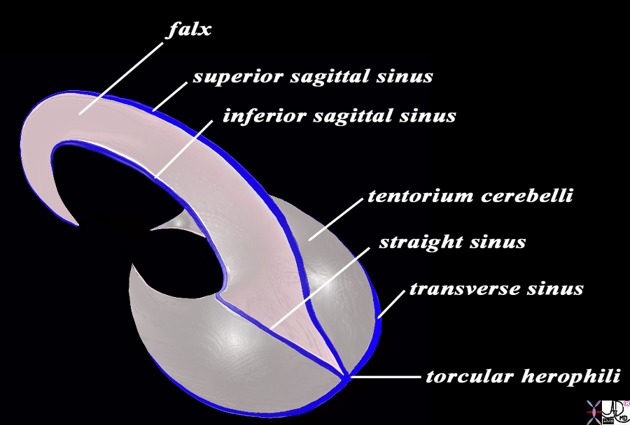
In this diagram the falx and the tentorium are exemplified together with their relationships to the dural sinuses. The image is drawn in a semisagittal plane showing the basic plan of the dural sinuses and relationship to the tentorium and falx. Note the sickle shaped falx posterior end having a greater craniocaudad dimension than the anterior end. The curved anterior aspect enables the falx to attach to the crista galli anteriorly as it proceeds under the frontal lobe. The tentorium cerebelli divides the brain into supratentorial and infratentorial structures. They are closed posteriorly and open anteriorly allowing the midbrain to pass through into the posterior fossa In the midline the straight sinus is enclosed and posteriorly the torcular herophili is present The superior sagittal sinus runs on the superior aspect of the falx and abuts the vertex of the skull. It usually empties into the right transverse sinus. The inferior sagittal sinus runs on the inferior aspect of the falx and empties into the straight sinus which in turn tends to empty into the left transverse sinus The transverse sinuses in turn empty into the sigmoid sinuses which exit the skull to enter the internal jugular veins. Courtesy Ashley Davidoff MD Copyright 2010 All rights reserved 98057b09.9sL |
Venous Phase of Arteriogram Showing the Venous Drainage of the Brain |
|
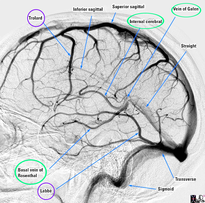
The diagram shows a venogram of the dural sinuses and highlighting the veins of the brain in lateral projection. The superficial veins (ringed in purple) consist of superior and inferior cerebral veins (not labeled). A branch of the inferior cerebral vein called the middle cerebral vein (not shown) provides collateral (vein of Trollard (purple ring)) that connects the inferior cerebral vein with the superior system by utilizing one of the superior cerebral veins. The vein of Labbe connects the middle cerebral vein with the transverse sinus . The common vein for the deep system (ringed in green) is the vein of Galen which empties into the straight sinus. It receives vessels from the internal cerebral vein and the vein of Rosenthal (both ringed in green) Image Courtesy Elisa Flower MD and Alex Norbash MD Copyright 2010 All rights reserved 97627b01.81s |
 The Dural Sinuses in Axial Projection Superimposed on the Skull Base The Dural Sinuses in Axial Projection Superimposed on the Skull Base |
|
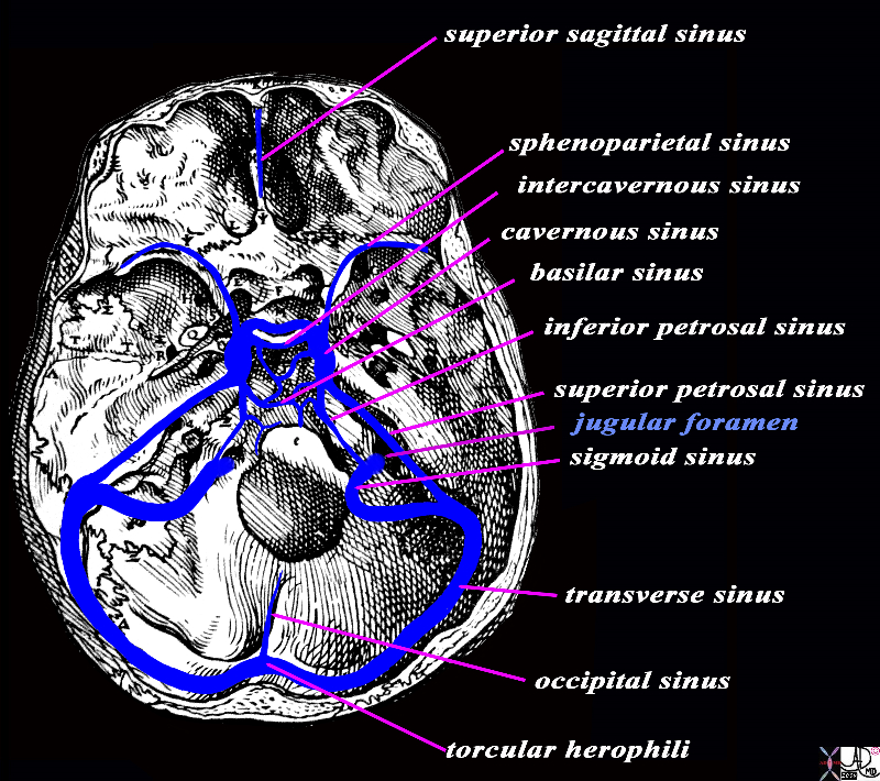
The diagram shows the dural sinuses and venous drainage of the brain in axial plane. The internal jugular vein is the final pathway through the jugular foramen (light blue text) It receives blood from the sigmoid sinus which drains the transverse sinus. The sigmoid sinus drains the inferior petrosal sinus, while the junction of the sigmoid to the transverse receives the superior petrosal sinus. At the base of the brain near the clivus the sinuses are a complex maze of interconnecting vessels with the epicenter being the cavernous sinuses. They connect the superior and inferior petrosal sinuses, basilar sinuses, intercavernous sinus, and sphenoparietal sinus. The pair of transverse sinuses are confluent with the occipital sinus and the straight sinus (not shown) at the confluens (torcular herophili). The superior sagittal sinus which runs on the superior aspect of the falx curves around by the frontal lobe and attaches to the crista galli as the anterior most sinus. Davidoff Art rendered with Vesalius skull -Vesalius image is in the public domain because its copyright has expired. 98058b06b.8s |
 A-P Venous Phase of an Arteriogram Showing the Large Dural Sinuses and Some of the Veins A-P Venous Phase of an Arteriogram Showing the Large Dural Sinuses and Some of the Veins |
|
The venous of an arteriogram in the A-P projection is shown revealing the cerebral veins draining into the superior sagittal sinus, some of which have pockets of arachnoid granulations protruding into the dural sinuses. The transverse sinus sigmoid sinus and internal jugular vein are easily recognised. The inferiorly positioned basal vein of Rosenthal and thalamostriate veins are more difficult to appreciate and the vein of Galen cannot be definitely identified in this view. Image Courtesy Ashley Davidoff MD Copyright 2010 49424b01 |
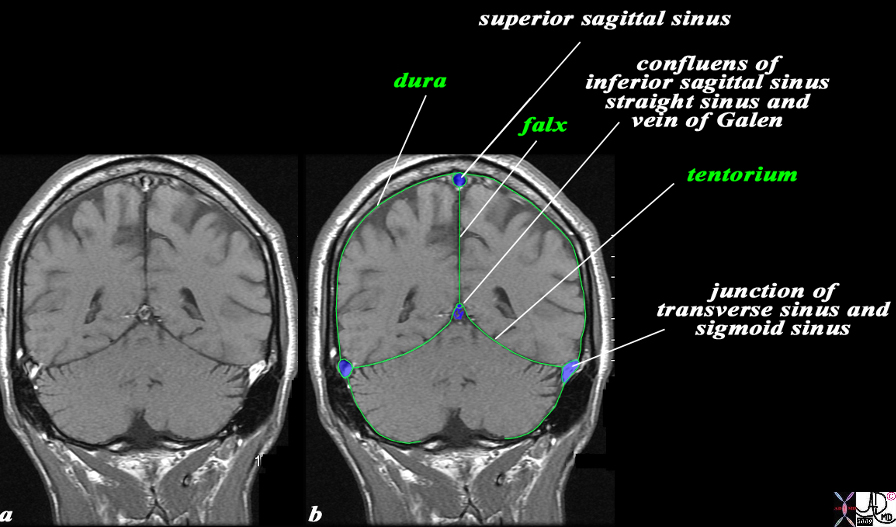 The Relationship of the Dural Sinuses and The Dura in the Coronal Plane The Relationship of the Dural Sinuses and The Dura in the Coronal Plane |
|
The overlay outlines the dural sinuses (blue) and the dura (green) in this coronal T1 weighted normal MRI sequence. The peripheral dural sinuses include the superior sagittal sinus, the laterally placed junction of the transverse sinus and straight sinus, and the confluens of the inferior sagittal sinus which runs on the inferior surface of the falx and the straight sinus which lies on the posterior and superior surface of the tentorium overlying the cerebellum. The dura is outlined in green and it splits to enclose the dural sinuses and also forms the tentorium. The dura splits at the vertex to encompass the superior sagittal sinus and then extends down in the interhemispheric fissure to form the falx, splits again to encompass the inferior sagittal sinus anteriorly and straigt sinus posteriorly, and then spreads out to form the tentorium. The thin black T1 dark layer outward of the dura is the inner cortical bone, and is followed by the T1 bright fat containing marrow of the skull. Next is the thin black T1 dark outer cortical bone, followed by a thick layer of T1 bright subcutaneous fat, and then a thin layer of isointense (to soft tissue) skin layer. Image Courtesy Ashley Davidoff MD Copyright 2010 All rights reserved 98170c04.9Ls |
CSF Drainage Arachnoid Granulations and Dural Sinuses
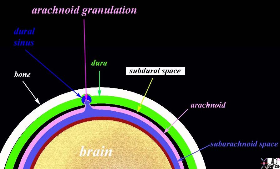 The Arachnoid Granulations and the Venous Sinuses The Arachnoid Granulations and the Venous Sinuses |
|
This diagram shows the morphology of the arachnoid granulation which is formed by the protrusion of the arachnoid membrane through the dura into the sinus. As it extends into the sinus it draws the subarachnoid space with it. The communication of the subarachnoid space and the arachnoid with the dural venous sinus allows the CSF to move from the subarachnoid space into the venous system. Also shown is the bone (white), dura (green), subdural space (black) arachnoid (pink) subarachnoid space (light blue) piamater (maroon) and the brain (cream colored) . Courtesy Ashley Davidoff MD Copyright 2010 98482b06.9 |
Applied Biology Diseases
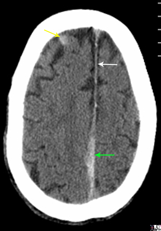 Acute Falcine and Frontal Subdural Hemorhage Acute Falcine and Frontal Subdural Hemorhage |
|
The non enhanced CTscan is from a 80 year old male on coumadin who sustained trauma to the head. There are two acute subdural hemorrhages. The first is a falcine subdural (green arrow) that has resulted in thickening irregularity and increased density of the falx near the vertex. This is contrasted to the normal falx anteriorly (white arrow) The second acute subdural is in the right frontal region (yellow arrow). Courtesy Ashley Davidoff MD Copyright 2010 89283.8sL |
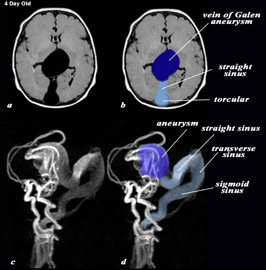 AVM, Enlarged Straight, Transverse and Sigmoid Sinuses and Vein of Galen Aneurysm AVM, Enlarged Straight, Transverse and Sigmoid Sinuses and Vein of Galen Aneurysm |
|
The MRI with a T1 weighted axial sequence (a,b) and with 3d TOF (c,d) shows a large aneurysm of the vein of Galen (dark blue (b,d) an enlarged straight sinus (light blue b) and enlarged draining transverse sinus and sigmoid sinus (c,d) The feeding vessels of the arteriovenous malformation (AVM) which is the cause of the aneurysm, seem to arise predominantly from the vertebro-basilar system and likely from the posterior cerebral artery, but the middle cerebral artery also appears to contribute to the AVM. Courtesy Philips Medical Systems 92432cL.9 |
—
References
Patel, M,R. Brain Imaging in Venous Sinus Thrombosis Emedicine