
The Common Vein Copyright 2010
Definition
The third ventricle represents the central cavity of the first brain vesicle. It is a narrow cavity in the diencephalon. It serves as a cushion to protect the brain from impacts that would otherwise damage the important structures contained in the diencephalon.
Size
The upper part is oval and its long axis (anteroposterior) measures 7 to 8 millimeters; the lower part measures 15 mm, and has the form of a triangle with its base directed upward.
Shape
It is shaped like a triangle whose apex is at the bottom.
A longitudinal groove, the sulcus of Monro, which extends from the foramen of Monro to the aqueduct of Sylvius, divides the walls of the ventricle into two parts, one superior and inferior.
It has four major recesses, named after the structures they are related to: a suprapineal recess, and immediately posterior to it a pineal recess. Continuing further down, the infundibular recess is present, and finally, the optical recess.
Position
It situated below the corpus callosum and between the thalami. Its floor is formed by the hypothalamus, the anterior wall by the lamina terminalis. It is middle and symmetrical. It communicates with the fourth ventricle through the aqueduct of Sylvius and with the lateral ventricles by the interventricular holes.
Character
Essentially a cavity whose walls are covered with ependyma, filled with CSF.
It is located between the thalamus and hypothalamus and contains cerebrospinal fluid. It communicates with the lateral ventricles though the interventricular foramina and the fourth ventricle through the cerebral aqueduct. The cerebral aqueduct is a thin tube between the third and fourth ventricles.
|
The vectors of the ventricular System Overlaid on the Brain |
|
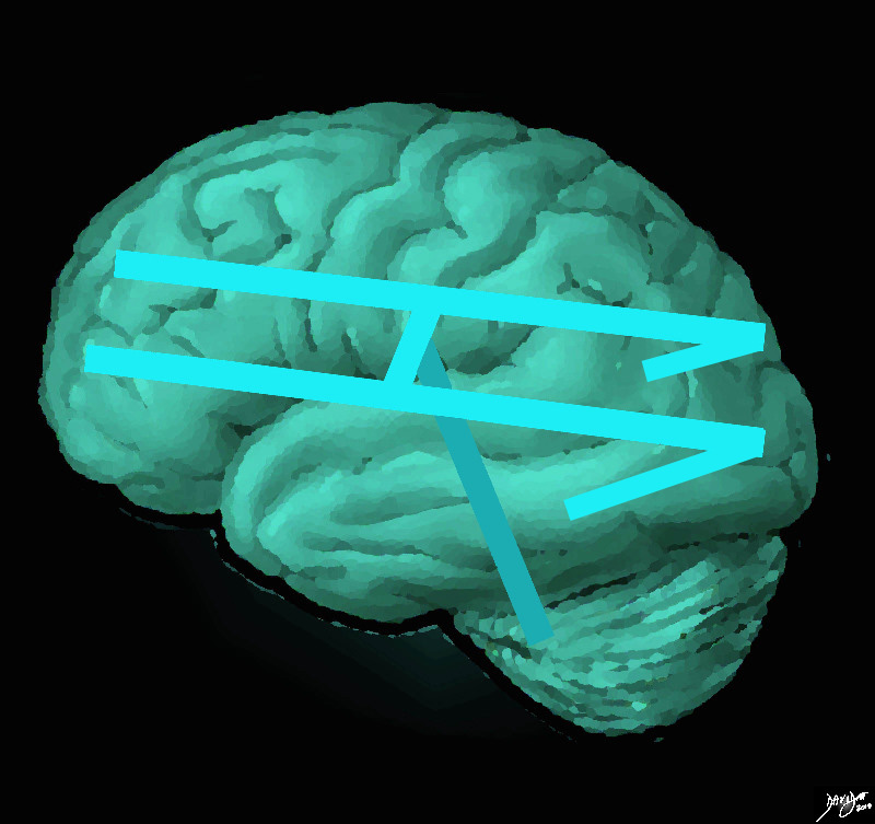
The ventricular system is overlaid on a drawing of the brain. Each limb of the horizontal component is situated in a cerebral hemisphere. The angled parts that extend inferiorly represent the temporal horns. The vertical limb includes the third and fourth ventricles Courtesy Ashley Davidoff MD copyright 2010 all rights reserved 94458e07.81s |
|
Sagittal View of the Ventricles |
|
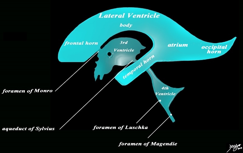
The diagram in the sagittal projection reveals the horizontal portion called the lateral ventricle. It is a paired structure that houses the frontal horn, body, and the vertical portion which is composed of the 3rd ventricle, cerebral aqueduct and the 4th ventricle. The lateral ventricle consists of the frontal horn, body, occipital horn, atrium and the temporal horn The foramen of Monro connects the lateral ventricles with the third ventricle. The paired foramina of Luschka are sitiuated anteriorly in the 4th ventricle and they allow CSF to circulate in the subarachnoid spaces. The foramen of Magendie is a single structure and is situated posteriorly and it also enables CSF to enter the subarachnoid space. Courtesy Ashley Davidoff MD copyright 2010 all rights reserved 94459b10b02.82s |
|
The Recesses |
|
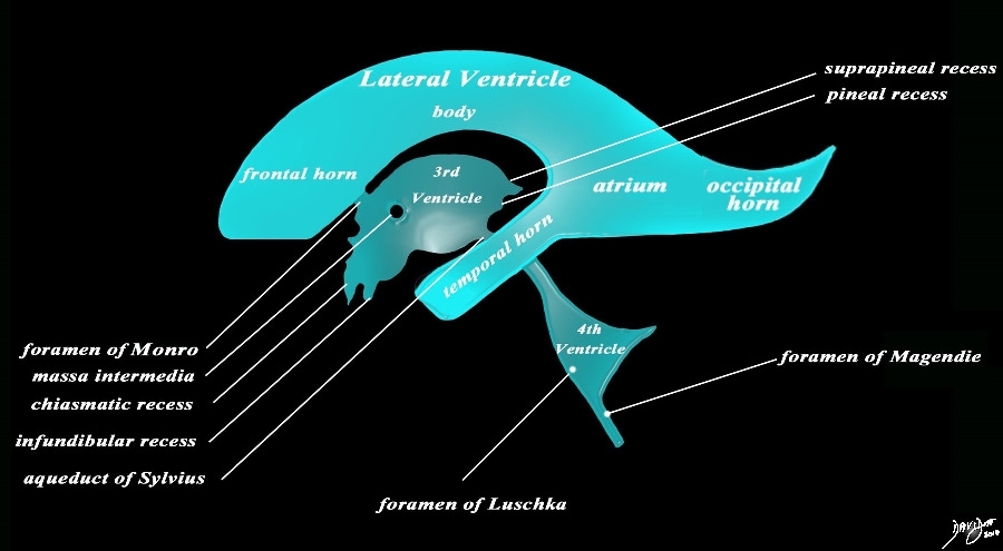
The diagram in the sagittal projection reveals the horizontal portion called the lateral ventricle. It is a paired structure that houses the frontal horn, body, and the vertical portion which is composed of the 3rd ventricle, cerebral aqueduct and the 4th ventricle. The lateral ventricle consists of the frontal horn, body, occipital horn, atrium and the temporal horn The foramen of Monro connects the lateral ventricles with the third ventricle. The paired foramina of Luschka are sitiuated anteriorly in the 4th ventricle and they allow CSF to circulate in the subarachnoid spaces. The foramen of Magendie is a single structure and is situated posteriorly and it also enables CSF to enter the subarachnoid space. code brain ventricles lateral ventricles frontal horn body occipital horn atrium temporal horn 3rd ventricle 4th ventricle foramen of Monro formamen of Magendie Foramen of Luschka anatomy normal neuroanatomy diagram conceptual diagram structure principles Davidoff Art Courtesy Ashley Davidoff MD copyright 2010 all rights Courtesy Ashley Davidoff MD copyright 2010 all rights reserved 94459b10b02.82s |
 Anatomic Specimen in Coronal Plane Anatomic Specimen in Coronal Plane
3rd Ventricle (3V) |
|
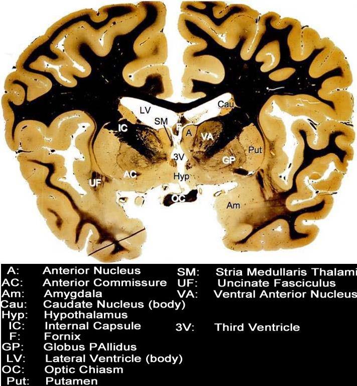
This coronal section through the forebrain reveals the more superficial cortical structures that include the frontoparietal part of the cerebral hemispheres and the temporal pole(TP). In the deeper portions of the brain and centrally positioned the corpus callosum (unlabelled white matter (black) that crosses the midline is appreciated. The fornix (F) is also seen as a midline structure just below the corpus callosum. The foramina of Munro (unlabelled) lead into the 3rd ventricle (3V). The hypothalamus (Hyp) follows and then the optic chiasm (OC). Alongside these central deep structures and lateral to the central structures, starting superiorly, the lateral ventricles (LV) are seen as two symmetrically positioned white structures. The anterior nucleus (A) and stria medullaris thalami (SM) lie alongside the third ventricle. Lateral to these structures and starting superiorly the caudate nucleus body (Cau) ventral anterior nucleus (VAN) internal capsule (IC) globus pallidus(GP), putamen (Put) amygdala (Am) and uncinate fasciculus (UF) are noted. Courtesy Department of Anatomy and Neurobiology at Boston University School of Medicine Dr. Jennifer Luebke , and Dr. Douglas Rosene 97346.C7.7L01 |
 Third Ventricle Third Ventricle
Coronal Plane STIR MRI |
|
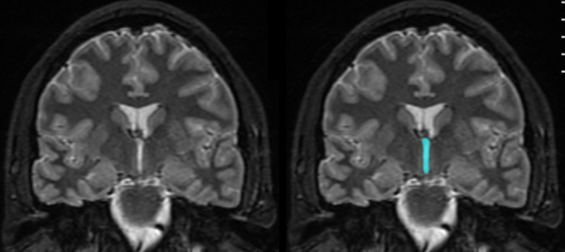
The third ventricle is demonstrated in this coronal MRI using a STIR sequence. It is easily recognized as a relatively narrow capital ” I ” shaped midline structure. It is connected to the lateral ventricles by the foramina of Munro (not obvious on these images). The temporal lobes are still apparent inferiorly at this plane. At this level some of the components of the basal ganglia can be evaluated below the lateral ventricles and lateral to the third ventricle Courtesy Ashley DAvidoff MD copyright 2010 all rights reserved 89735c01.8s |
|
Foramina of Munro Defining the Posterior Border of the Frontal Horns |
|
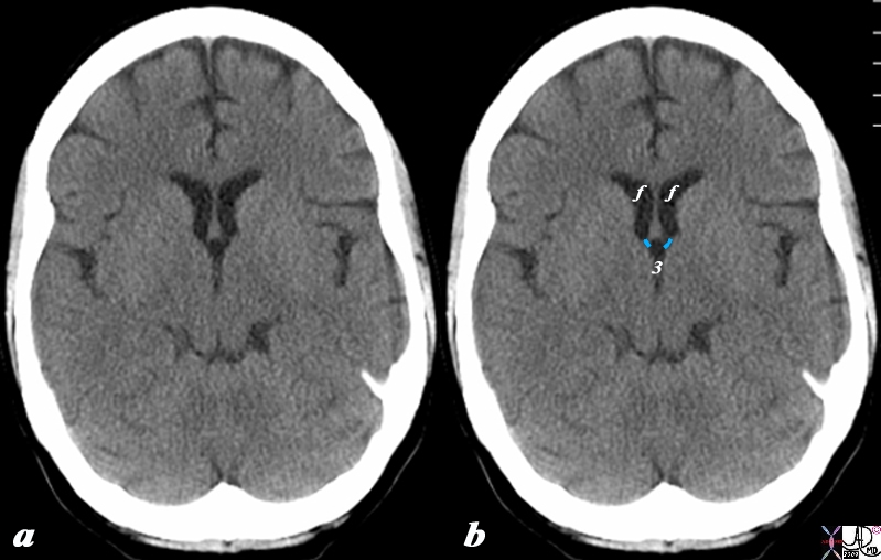
The CT scan in axial projection reveals the frontal horns (f) connecting with the 3rd ventricle (3) via the small foramina of Munro (overlaid in light blue). The posterior border of the frontal horns is defined by the foramen of Munro on either side. Courtesy Ashley Davidoff MD Copyright 2010 All rights reserved 98169cL.8s |
|
Normal Asymmetry of the Lateral Ventricles Symmetry of the Small Foamina of Munro |
|
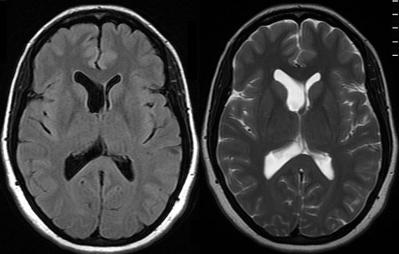
The MRI shows a FLAIR and a T2 weighted image revealing normal asymmetric development of the lateral ventricles in a 41 year old female. Note however the two symmetric small channels representing the foramina of Munro which are not affected by the asymmetry. Courtesy Ashley Davidoff MD Copyright 2010 89044c |
 Relationship of the Hypothalamus with the Third Ventricle Relationship of the Hypothalamus with the Third Ventricle |
|
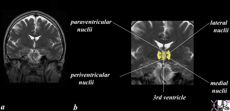
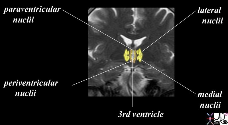
The hypothalamus is centered in the midline around the third ventricle. The coronal image of a T2 weighted MRI shows the third ventricle in the midline (white) immediately surrounded by a the thinnest layer of paraventricular component, which in turn is surrounded by a slighly thicker layer of periventricular component (orange followed laterally by a pair of medial nuclii and then a single larger lateral component. The image below is a magnified version to enable you to appreciate the two inner layers better. \ Courtesy Ashley Davidoff MD copyright 2010 60528.8c11.8s |
 Relations of the Third ventricle in Coronal Projection Relations of the Third ventricle in Coronal Projection
MRI T2 Weighted Image |
|
The slightly magnified view of the coronal image of a T2 weighted MRI shown above again shows the third ventricle in the midline (white). The thinnest inner layer is a mere pencil thin yellow line (paraventricular layer) and is barely seen even in this magnified view. The periventricular layer surrounds it. (orange) Courtesy Ashley Davidoff MD copyright 2010 60528.8c09.8s |
|
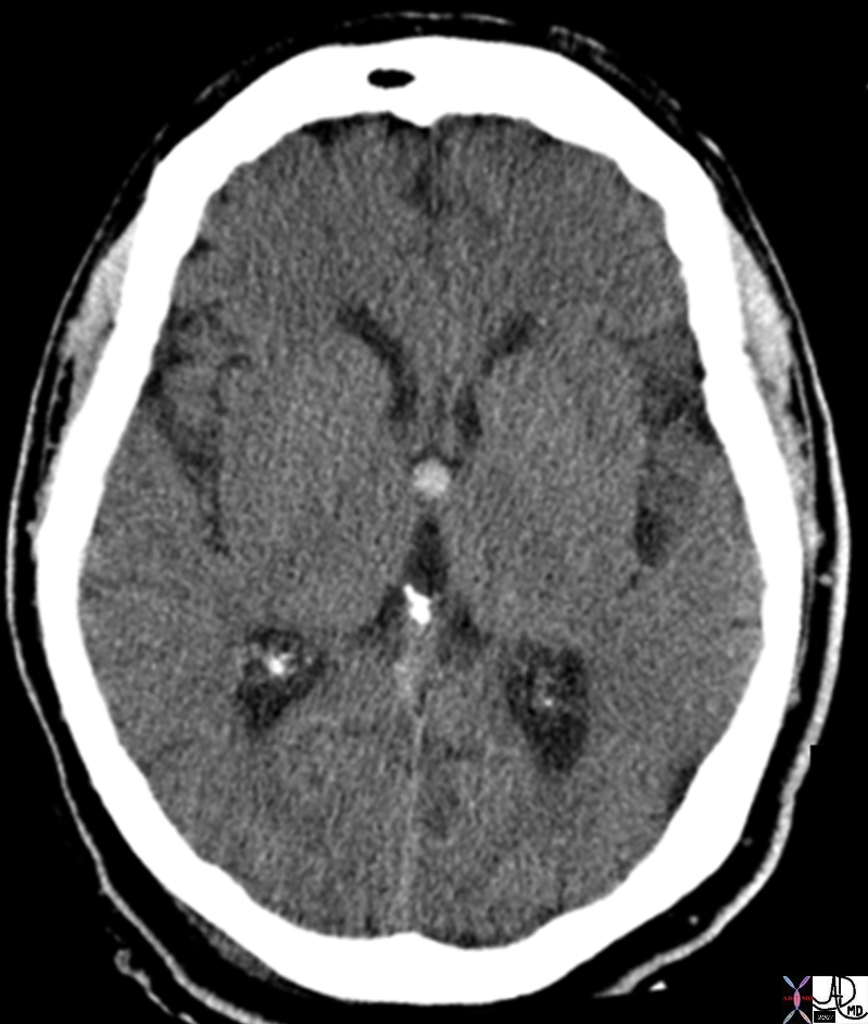
Colloid Cyst of Third Ventricle – Indication for Surgery of Benign Tumor |
| The CT scan is from a young adult female who presented with a history of trauma and a subcentrimetre hyperdense lesion identified was an incidental finding.On the non contrast CT a smooth round hyperdense nodule is noted in the region of the foramen of Munro and the anterior aspect of the third ventricle. These findings are consistent with a diagnosis of a colloid cyst.
Pathologically these lesions are benign and filled with gelatinous material and cholesterol crystals which are responsible for the hyperdensity. They can rupture or cause hydrocephalus. Image Courtesy Ashley Davidoff MD 70045.800 |
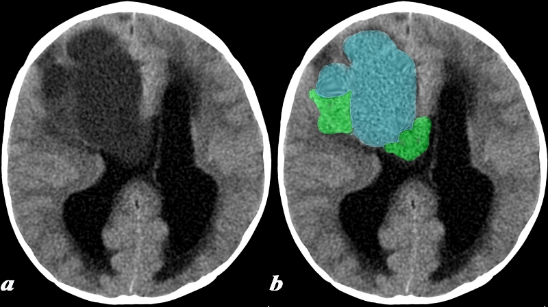
 Choroid Plexus Papilloma of the Third Ventricle and Hydrocephalus Choroid Plexus Papilloma of the Third Ventricle and Hydrocephalus |
|
The coronal MRI shows a gadolinium enhanced T1 weighted image (a,b) with black CSF in the dilated ventricles and a T2 weighted image with bright CSF (c,d). A large mass in the third ventricle (overlaid ingreen) dominates the study and is complicated by severe hydrocephalus. The mass was a choroid plexus papilloma of the third ventricle. In patients with brain tumor one of the causes of headache is hydrocephalus. This is an extreme case of hydrocephalus. Courtesy James Donnelly MD 23223c01 |
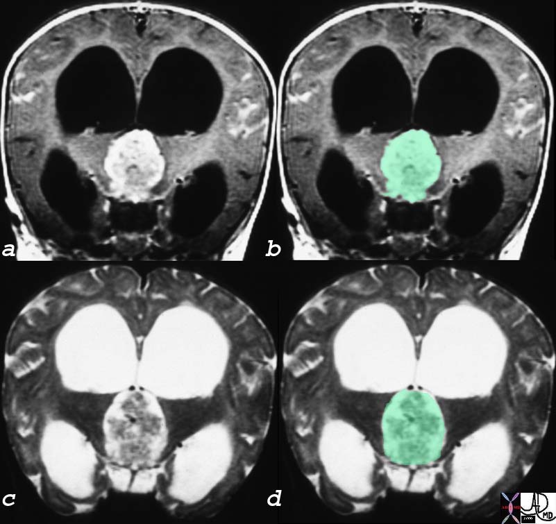
 Desmoplastic Infantile Ganglioganglioma Causing Obstruction of the Third Ventricle Desmoplastic Infantile Ganglioganglioma Causing Obstruction of the Third Ventricle |
|
This 17 month old boy presented with vomiting. CT: (a,b) A noncontrast head CT was obtained and demonstrates a predominantly cystic lesion (blue) with more solid components (green) at the periphery. Notice the enlargement of the lateral ventricles which is due to obstructive hydrocephalus caused by obstruction at the level of the third ventricle (not included on this image). These findings are consistent with a desmoplastic infantile ganglioglioma (DIG) Image Courtesy Elisa Flower MD and Asim Mian MD 97641c.8 |