The Common Vein Copyright 2010
Definition
The dura mater (pachymenix) or “tough mother” is a thick, tough collagen membrane, which is found as a double layer as the the outer third layer of the meninges. It is comprised of an outer and inner layer. The outer latyer formas the inner periosteal layer of th the skull, and the inner layer connects with the arachnoid layer of the meninges. The two layers function as a single unit but they spit to enclose and in fact form the form dural venous sinuses, into which the cerebral veins drain (
The dural connections to the skull and the arachnoid meninges enable two spaces; the epidural space and the subdural space. These potential spaces are appreciated most commonly after bleeding occurs folowing trauma.
|
General Organization of the Meninges |
|
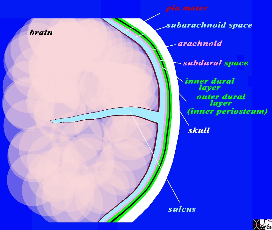
The coronal drawing reveals the three layers of the brain. The inner layer (maroon) is the pia mater and it is in intimate contact with the brain and faithfully follows the sulci and gyri. The second layer is the arachnoid (pink) which is a slightly thicker membrane and follows the pia in a general fashion but does not extend into the sulci. It is intimately attached to the inner layer of the dura (bright green) The space between the pia and the arachnoid is the subarachnoid space, and it is in this space that the CSF is present and surrounds the brain. The next layer is the dura which is a double layer. The inner layer (bright green) is intimately attached to the arachnoid and the outer layer (also bright green) is attached to the bone and functions as the inner periosteum. There is a potential space between the arachnoid (pink) and the inner layer of the dura (green). This space is called the subdural space. (combination pink and green) The CSF (light blue) is seen in the subarachnoid space and in the lateral ventricles (gray blue) Image Courtesy Ashley Davidoff MD Copyright 2010 All rights reserved 71422.800b02g05.91s |
|
Dura and Arachnoid and Subarachnoid Space MRI T1 Weighted Coronal View |
|
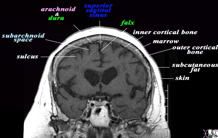
The coronal T1 weighted MRI reveals demonstrates some of the anatomical features of the meninges and their relations The sulci are seen as T1 dark CSF containing fissures in the brain substance. The pia is too thin to see but would be adherent to the brain surface. The space that contains the CSF is called the subarachnoid space (light blue). The very fine T2 bright membrane seen on the surface of the brain is a combination of arachnoid (pink) adherent to the two layers of the dura (green). The outer layer of the dura acts as the inner periosteum and is adherent to the skull. The dura splits at the vertex to encompass the superior sagittal sinus and then extends down in the interhemispheric fissure to form the falx. The thin black T1 dark layer after the arachnoid and dura is the inner cortical bone, and is followed by the T1 bright fat containing marrow of the skull. Next is the thin black T1 dark outer cortical bone, followed by a thick layer of T1 bright subcutaneous fat, and then a thin layer of isointense (to soft tissue) skin layer. Image Courtesy Ashley Davidoff MD Copyright 2010 All rights reserved 71422.800bL01.9 |
|
Dura and Arachnoid and Subarachnoid Space MRI T1 Weighted Coronal View |
|
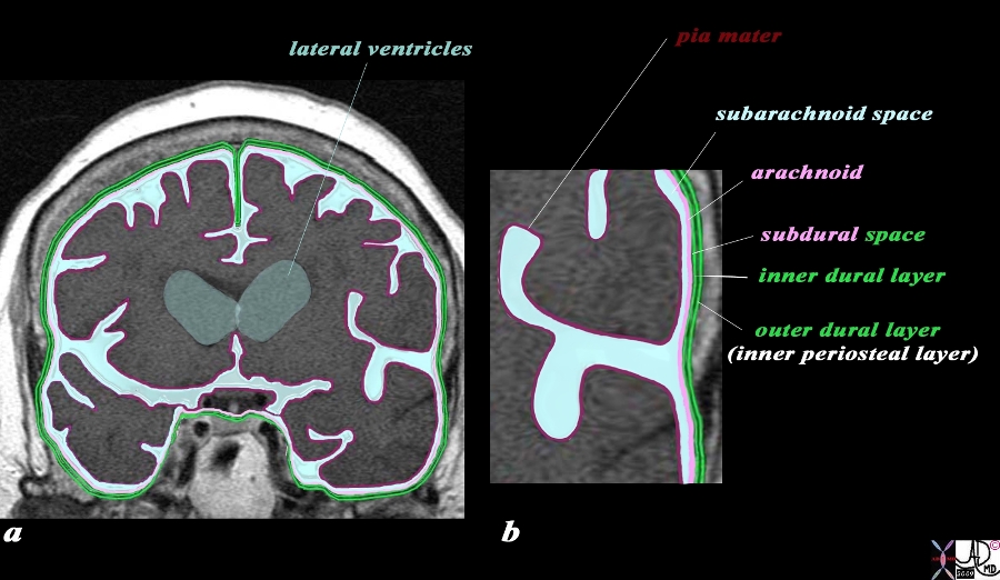
The coronal image of the brain using T1 weighted MRI sequence reveals the three layers of the brain. The inner layer (maroon) is the pia mater and it is in intimate contact with the brain and faithfully follows the sulci and gyri. The second layer is the arachnoid (pink) which is a slightly thicker membrane and follows the pia in a general fashion but does not extend into the sulci. It is intimately attached to the inner layer of the dura (bright green) The space between the pia and the arachnoid is the subarachnoid space, and it is in this space that the CSF that surrounds the brain is housed. The next layer is the dura which is a double layer. The inner layer (bright green) is intimately attached to the arachnoid and the outer layer (also bright green) is attached to the bone and functions as the inner periosteum. There is a potential space between the two layers of the dura. The CSF (light blue) is seen in the subarachnoid space and in the lateral ventricles (gray blue) Image Courtesy Ashley Davidoff MD Copyright 2010 All rights reserved 71422.800b04c01.9s |
|
The Relationship of the Dural Sinuses and The Dura in the Coronal Plane |
|
The overlay outlines the dural sinuses (blue) and the dura (green) in this coronal T1 weighted normal MRI sequence. The peripheral dural sinuses include the superior sagittal sinus, the laterally placed junction of the transverse sinus and straight sinus, and the confluens of the inferior sagittal sinus which runs on the inferior surface of the falx and the straight sinus which lies on the posterior and superior surface of the tentorium overlying the cerebellum. The dura is outlined in green and it splits to enclose the dural sinuses and also forms the tentorium. The dura splits at the vertex to encompass the superior sagittal sinus and then extends down in the interhemispheric fissure to form the falx, splits again to encompass the inferior sagittal sinus anteriorly and straigt sinus posteriorly, and then spreads out to form the tentorium. The thin black T1 dark layer outward of the dura is the inner cortical bone, and is followed by the T1 bright fat containing marrow of the skull. Next is the thin black T1 dark outer cortical bone, followed by a thick layer of T1 bright subcutaneous fat, and then a thin layer of isointense (to soft tissue) skin layer. Image Courtesy Ashley Davidoff MD Copyright 2010 All rights reserved 98170c04.92Ls |
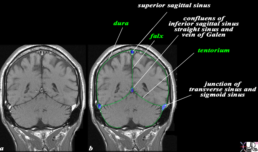
The dura mater also has three major reflections. The falx (sickle) cerberi, is a sickle shaped reflection of dura that projects between the cerebral hemishpheres in the interhemispheric fissure vertically, and incompletely divides the two cerebral hemispheres. It has attachments to the ethmoid bone (crista galli) anteriorly. Posteriorly, it attaches to another dural fold, the tentorium (tent) cerebelli.
The tentorium is a horizontal dural reflection which divides the cerebellum and divides the brain into a supratentorial compartment and an infratentorial compartment. The tentorium attaches to the occipital bone of the skull posteriorly. The anterior part of the tentorium is open allowing the brainstem to passes. This opening is called the tentorial notch.
Applied Biology
Fractures – usually tear the dura
Subdural hematoma is the most common dural trumatic injury occurring in the elederly since the brain is shrunken and the vessels are therefore relativley stretched and with trauma they bleed under the dura either immadiately or even subsequently.
The hematom occurs between the inner layer of the dura and the arachnoid.
A chronic subdural becomes hyperosmolar and th collection slowly draws in fluid and over a few weeks, gets larger and the patient may become unconscious as a result in the subacute phase.
One of the first surgical operations was the relief of subdural hematoma
|
Subacute Subdural hematoma with Enhancing and Thickened Arachnoid CT scan |
|
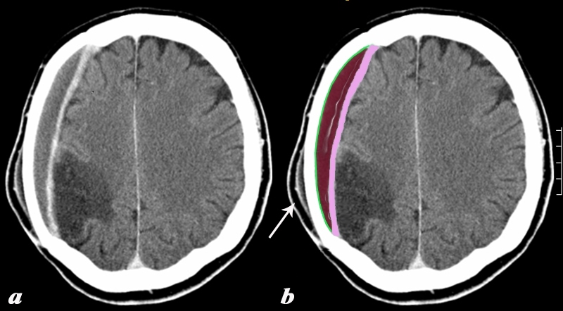
The CTscan with contrast shows a collection that conforms to the shape of the skull with an enhancing meningeal layer. The right parietal collection is a subdural hematoma (SDH) with the outer border conforming to the shape of the skull and lined by the two layers of dura (green) and the inner border thickened and enhancing lined by the arachnoid (pink). The arachnoid is unusually thick in this case suggesting some complexity to the subdural. In the appropriate clinical setting this could be an infected subdural. A subacute subgaleal subcutaneous hematoma is seen in the soft tissues overlying the SDH (white arrow). It also has a slightly enhancing outer layer In addition, as an unrelated abnormality, there is a chronic infarct of the right parietal lobe characterized by a hypodense triangular defect, with loss of gray white matter interface, all consistent with encephalomalacia. Courtesy Ashley Davidoff MD Copyright 2010 89273c03 |
 Bilateral Subdural Hematomas Bilateral Subdural Hematomas |
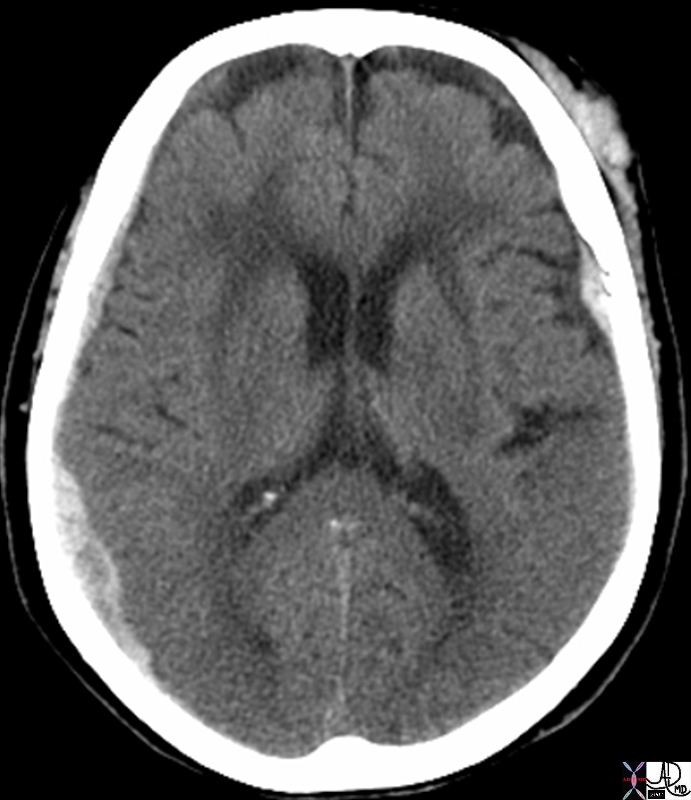
| In this patient the non contrast CTscan reveals small bilateral subdural hematomas . In the left frontal region there is a subgaleal hematoma and underlying the hematoma there is a small hyperdense subdural hematoma in the region of the Sylvian fissure.
A contracoup injury manifesting also as a subdural hematoma is seen in the right posterior parietal region. There are linear hyperdensities overlying the frontal lobes bilaterally that may represent small acute hemorrhages as well. 70000.800 skin brain cerebrum cerebral subcutaneous subdural contra coup fx acute blood fx density dense subdural hematoma subcutaneous hematoma trauma radiologists and detectives CTscan Davidoff MD |
|
Acute Falcine and Frontal Subdural Hemorhage |
|
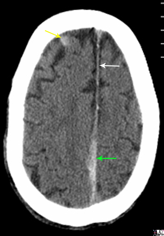
The non enhanced CTscan is from a 80 year old male on coumadin who sustained trauma to the head. There are two acte subdural hemorrhages. The first is a falcine subdural (green arrow) that has resulted in thickening irregularity and increased density of the falx near the vertex. This is contrasted to the normal falx anteriorly (white arrow) The second acute subdural is in the right frontal region (yellow arrow). Courtesy Ashley Davidoff MD Copyright 2010 89283.8sL |
The epidural hematoma also occurs as a result of blunt trauma to skull usually in younger patients and is more commonly an arterial bleed. A classical syndrome of momentary loss of conciousness manifesting clinically as a concussion, the patient becomes lucid again but with ongoing bleeding they become unconscious again as the hematoma expands.
The hematoma occurs beneath the skull and pushes the inner layer of the periostem (outer dural layer) off the skull resulting in both layers of the dura and the arachnoid being on the inner surface of the hematoma.
|
Epidural Hematoma Skull Fracture |
|
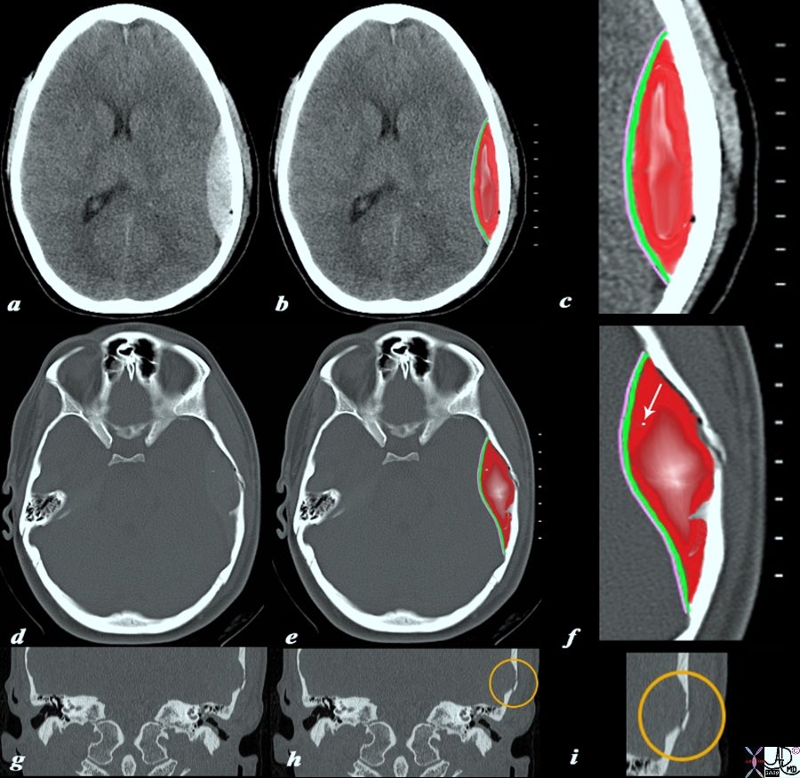
The CTscan without contrast is from a 19 year old male who sustained head trauma and who is complaining of a severe headache. The scan shows a high density lenticular shaped hematoma (a,b,c),. The right parietal collection is an extradural or epidural hematoma, with the outer border conforming to the shape of the skull and lined by the bone) and the inner border lined by two layers of dura (green c,f) and the arachnoid (pink c,f). There are bubbles of air scattered along the inner bone surface (black bubbles best seen on the magnified views (c, f) a tiny fragment of bone (white arrow in f) but seen on images d, and e as a white speck. The fracture of the temporal bone is ringed in orange on the coronal reconstructions of the axial views (h,i) Courtesy Ashley Davidoff MD Copyright 2010 98056c04.9s |