
The Common Vein Copyright 2010
Definition
An intracranial lipoma is characterized as a congenital anomlay. They are thought to be caused by an abnormal persistence and differentiation of the mesenchymal derivative of the neural crest called the meninx primitiva. Lipomas are most frequently seen in the pericallosal region but can also be found in the suprasellar and quadrigeminal plate cisterns. They are most commonly associated with hypogenesis or agenesis of the corpus callosum.
Pathologically, intracranial lipomas are identical to adipose tissue found elsewhere in the body.
Clinically, lipomas are usually asymptomatic. Seizures and mental disorders are often present as well but these may be associated with the concomitant anomalies.
Diagnosis can be made by classic imaging findings.
Imaging includes CT, MRI, and potentially ultrasound when performed on a neonate through an open fontanel. On ultrasound, lipomas appear as hyperechoic masses. On CT, they follow the low density of fat. Lipomas in association with the corpus callosum can be partially calcified. On MRI, they follow the signal intensity of fat, being T1 and T2 hyperintense and drop out in signal on fat suppressed sequences. They generally do not enhance.
Treatment is reserved for symptomatic patients, for example if a lipoma in the quadrigeminal cistern is large enough to cause hydperocephalus, but otherwise there is no treatment.
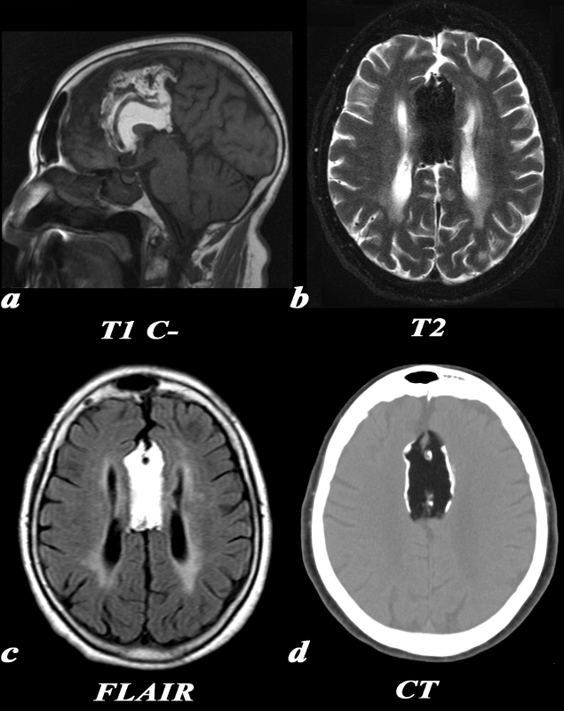
Pericallosal Lipoma and Partial Agenesis of the Corpus Callosum |
|
This 65 year old female was noted to have a large pericallosal lipoma. MRI, T1 (a): The large pericallosal lipoma is T1 hyperintense and follows the signal intensity of fat. There is partial agenesis of the corpus callosum, with only the genu being visualized on this midline sagittal image. Also note the anterior cerebral arteries coursing directly through the lipoma T2 (b): This T2 weighted image is obtained using a technique which suppresses fat. The signal within the lipoma has been suppressed causing it to be hypointense. FLAIR (c): On this FLAIR sequence, again note that the signal in the lesion follows that of fat signal. Also note the parallel configuration of the lateral ventricles consistent with partial agenesis of the corpus callosum. CT (d): On CT, the low density matches that of the subcutaneous fat. This lipoma has peripheral calcification. . The diagnosis is consistent with a pericallosal lipoma with peripheral calcification and partial agenesis of the corpus callosum Image Courtesy Elisa Flower MD and Asim Mian MD 97660c.8 |