Subarachnoid Hemorrhage
The Common Vein Copyright 2010
Definition
Subarachnoid hemorrhage is bleeding into the subarachnoid space – the area between the arachnoid membrane and the pia mater.
Ruptured saccular (berry) aneurysm is the most common cause. Trauma and AV malformation are other common causes. Common sites of the origin of the hemorrhage are at the junction of anterior communicating artery with anterior cerebral artery, the junction of posterior communicating artery with the internal carotid artery and at the bifurcation of the MCA
Patients present with sudden, severe (often excruciating) headache in the absence of focal neurologic symptoms. Half of the patients can be present after a sudden and transient loss of consciousness. Vomiting (intracranial hypertension) and signs of meningeal irritation, such as nuchal rigidity, and photophobia develop as the hemorrhage expands. EKG changes are common immediately after the hemorrhage, leading to sometimes potentially fatal arrhythmias, though benign in their most part.
Noncontrast CT’s identify the majority of SAH’s, by showing blood, hyperdense, in the subarachnoid space. It can be falsely negative in up to 10% of cases. When clinical suspicion is high, a LP should be performed, as it will be diagnostic, revealing blood and xanthochromia. It can also provide an estimate of the amount of blood extravagated into the cisterns and ventricles.
A cerebral angiography should follow once the diagnosis is confirmed, as it will identify the location of the bleed, and can also offer a possibility for treatment through surgical clipping.
Structural Considerations

The Subarachnoid Space Between the Pia and Arachnoid (light blue) |
|
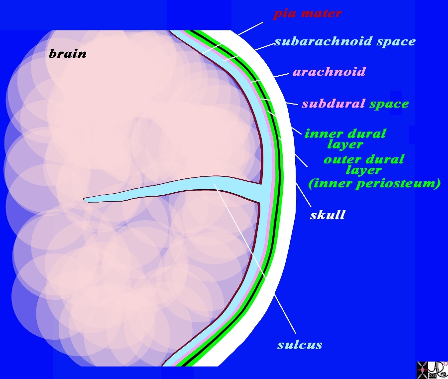
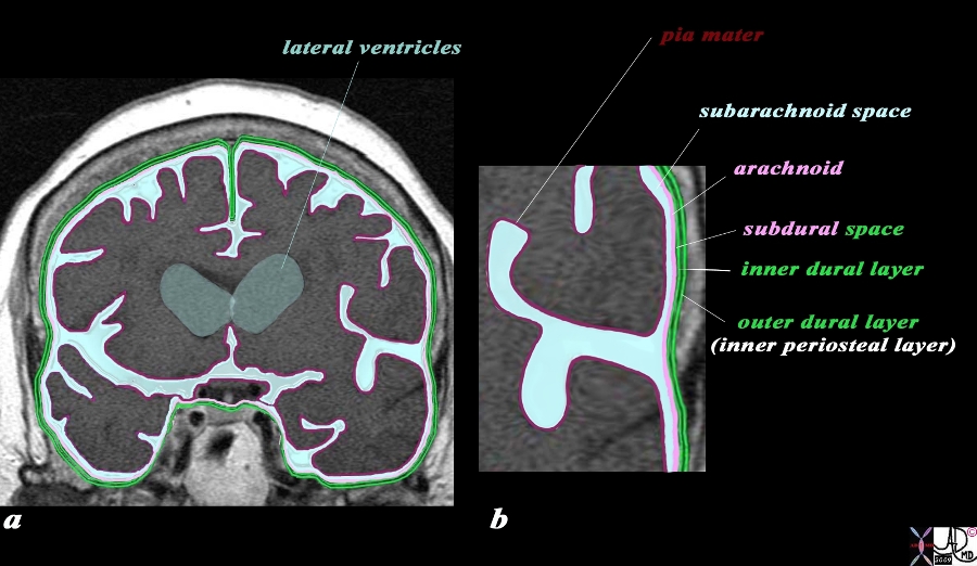
The coronal drawing reveals the three layers of the brain. The inner layer (maroon) is the pia mater and it is in intimate contact with the brain and faithfully follows the sulci and gyri. The second layer is the arachnoid (pink) which is a slightly thicker membrane and follows the pia in a general fashion but does not extend into the sulci. It is intimately attached to the inner layer of the dura (bright green) The space between the pia and the arachnoid is the subarachnoid space, and it is in this space that the CSF is present and surrounds the brain. The next layer is the dura which is a double layer. The inner layer (bright green) is intimately attached to the arachnoid and the outer layer (also bright green) is attached to the bone and functions as the inner periosteum. There is a potential space between the arachnoid (pink) and the inner layer of the dura (green). This space is called the subdural space. (combination pink and green) The CSF (light blue) is seen in the subarachnoid space and in the lateral ventricles (gray blue) c Courtesy Ashley Davidoff MD Copyright 2010 All rights reserved 71422.800b02g06.91s |
|
Dura and Arachnoid and Subarachnoid Space MRI T1 Weighted Coronal View |
|
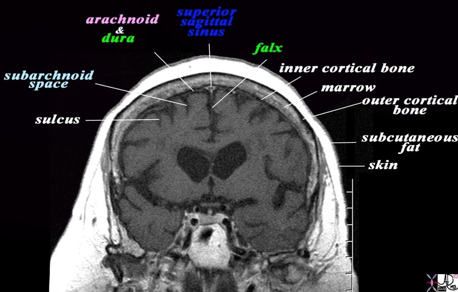
The coronal T1 weighted MRI reveals demonstrates some of the anatomical features of the meninges and their relations The sulci are seen as T1 dark CSF containing fissures in the brain substance. The pia is too thin to see but would be adherent to the brain surface. The space that contains the CSF is called the subarachnoid space (light blue). The very fine T2 bright membrane seen on the surface of the brain is a combination of arachnoid (pink) adherent to the two layers of the dura (green). The outer layer of the dura acts as the inner periosteum and is adherent to the skull. The dura splits at the vertex to encompass the superior sagittal sinus and then extends down in the interhemispheric fissure to form the falx. The thin black T1 dark layer after the arachnoid and dura is the inner cortical bone, and is followed by the T1 bright fat containing marrow of the skull. Next is the thin black T1 dark outer cortical bone, followed by a thick layer of T1 bright subcutaneous fat, and then a thin layer of isointense (to soft tissue) skin layer. Image Courtesy Ashley Davidoff MD Copyright 2010 All rights reserved 71422.800bL01.9 |

Dura and Arachnoid and Subarachnoid Space MRI T1 Weighted Coronal View |
|
The coronal image of the brain using T1 weighted MRI sequence reveals the three layers of the brain. The inner layer (maroon) is the pia mater and it is in intimate contact with the brain and faithfully follows the sulci and gyri. The second layer is the arachnoid (pink) which is a slightly thicker membrane and follows the pia in a general fashion but does not extend into the sulci. It is intimately attached to the inner layer of the dura (bright green) The space between the pia and the arachnoid is the subarachnoid space, and it is in this space that the CSF that surrounds the brain is housed. The next layer is the dura which is a double layer. The inner layer (bright green) is intimately attached to the arachnoid and the outer layer (also bright green) is attached to the bone and functions as the inner periosteum. There is a potential space between the two layers of the dura. The CSF (light blue) is seen in the subarachnoid space and in the lateral ventricles (gray blue) Image Courtesy Ashley Davidoff MD Copyright 2010 All rights reserved 71422.800b04c01.9s |

Acute Subarachnoid Hemorrhage |
|
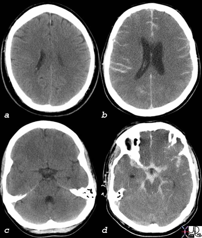
The CT scan of this 52 year male is without contrats and reveals findings consistent with a subarachnoid hemorrhage likely caused by rupture of an aneurysm. In image b high density blood is seen in the sulci and in the interhemispheric fissureand in image d hemorrhage is seen in the stellate shaped suprasellar cistern and in the interpeduncular cistern of the midbrain, ambient cisterns, and likely in the aqueduct. Courtesy Ashley Davidoff MD 76059c01 |

