Sarcoidosis of the Brain and Meninges
The Common Vein Coopyright 2010
Definition
Sarcoidosis is a disease characterized by the presence of noncaseating granulomas that can be present in multiple organs. It has an unknown cause, and it affects mostly the lungs and the lymph nodes in the mediastinum and hilar regions. The natural history is variable, as it sometimes can spontaneously resolve, but most of the times it is chronic and progressive, leading to (multi)organ failure and possibly death.
Being systemic, it can affect the neurological system (neurosarcoidosis) in up to 10% of these patients. Any part of this system can be involved; the base of the brain tends to be more frequent.
Pathologically, collections of epithelioid cells develop, being surrounded by a rim of lymphocytes (granuloma), with giant cell formation. Unlike tuberculosis, there is no caseum.
In terms of symptoms, non-specific complains are frequently present, due to its systemic nature – fever, weight loss, and fatigue. Peripheral mononeuropathies and polyradiculoneuropathy can be present, if nerves are affected. In fact, the most common symptom in neurologic involvement is unilateral facial nerve palsy. Due to compression in the CNS, seizures can occur, as well as corticospinal and cerebellar signs. The hypothalamus and pituitary gland can be involved, giving rise to hyperprolactinemia, diabetes insipidus and visual field changes. There can be aseptic meningitis. In terms of cranial neuropathy, usually the facial nerve is the most commonly affected, followed by the optic nerve and the trigeminal. In cranial neuropathy, besides the granulomatous infiltration, leptomeningeal fibrosis also plays an important role.
Diagnosis is clinical, along with presence in biopsy of sarcoid granulomas in the body.
Radiologically, central neurosarcoidosis can have multiple patterns, and that will depend on which components are affected. When there is meningeal or ependymal invasion, meningeal nodules, and most commonly a diffuse meningeal enhancement is seen, which is most obvious in the basal cisterns. In the brain parenchyma, if there is invasion, CT may show homogenous isodense or hyperdense nodule(s).
Patients with neurosarcoidosis, may respond well to corticosteroids; if resistant, immunosuppressive agents can be used, such as cyclosporine.

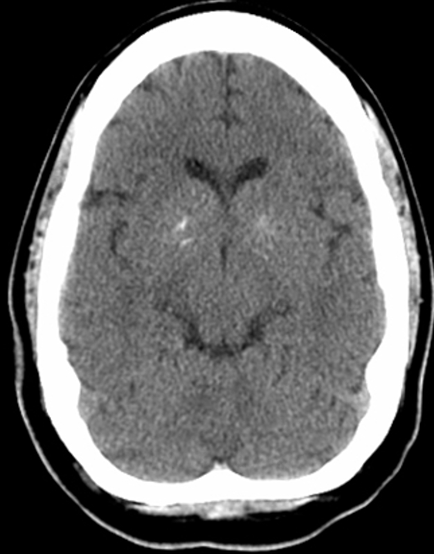
Basal Ganglia Calcification in a 29 year old with Sarcoidosis |
|
Calcification in the basal ganglia in the region of the globus pallidus is shown in axial projection in this CTscan of a 29 year old female with sarcoidosis. The scan is otherwise normal. It is likely that there is granulomatous involvement of the basal ganglia with sarcoidosis Courtesy Ashley Davidoff MD Copyright 2010 All rights reserved 89070.8 |
