Aqueduct of Sylvius – Cerebral Aqueduct
The Common Vein Copyright 2010
Definition
The aqueduct of Sylvius is the small channel that connnects the 3rd ventricle with the 4th ventricle. It is located in the midbrain and defines the border between the tectum and tegmentum of the midbrain. It is surrounded by periaqueductal gray matter and lies just posterior to the oculomotor nuccli.
|
Sagittal View of the Ventricles |
|
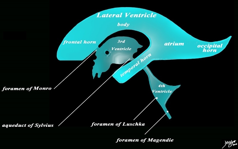
The diagram in the sagittal projection reveals the horizontal portion called the lateral ventricle. It is a paired structure that houses the frontal horn, body, and the vertical portion which is composed of the 3rd ventricle, cerebral aqueduct and the 4th ventricle. The lateral ventricle consists of the frontal horn, body, occipital horn, atrium and the temporal horn The foramen of Monro connects the lateral ventricles with the third ventricle. The paired foramina of Luschka are sitiuated anteriorly in the 4th ventricle and they allow CSF to circulate in the subarachnoid spaces. The foramen of Magendie is a single structure and is situated posteriorly and it also enables CSF to enter the subarachnoid space. Courtesy Ashley Davidoff MD copyright 2010 all rights reserved 94459b10b02.82s |
|
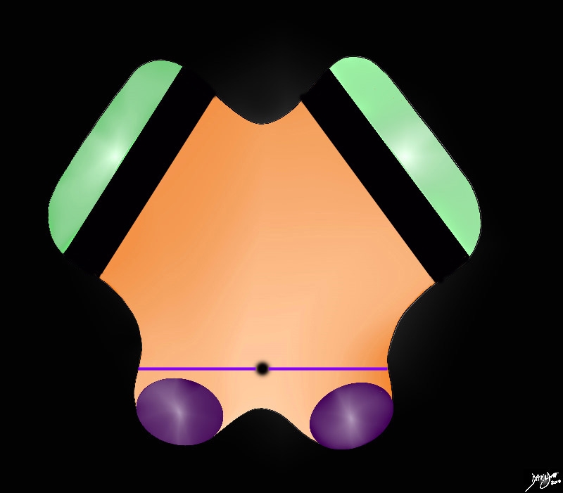
Aqueduct (black hole) The Border between the Tegmnetum and Tectum) |
|
The anterior border of the midbrain incorporates the cerbral peduncles(green), and the substantia nigra (black – just posterior to the peduncles. Between the substantia nigra and the aqueduct is an area of the midbrain called the tegmentum (floor of the midbrain) The posterior end of the midbrain is bordered by the colliculi (purple) in the tectum (roof) of the midbrain. Courtesy Ashley Davidoff MD copyright 2010 all rights reserved 94074b09b05b.83s |
|
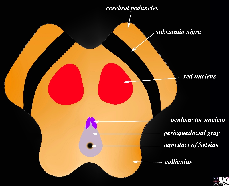
Aqueduct in Axial Projection Surrounded by periaqueductal Gray |
|
The midbrain in transverse plane illustrates the component structures, with the image reminiscent of the face of a baby pig.. Anteriorly the cerebral peduncles are followed by the substantia nigra, and the collilculi The anterior border of the midbrain incorporates the cerebral peduncles, and the substantia nigra (black – just posterior to the peduncles). Between the substantia nigra and the aqueduct is an area of the midbrain called the tegmentum (floor of the midbrain) Within the tegmentum other structures include red nuclii, oculomotor nuclii, periaquaductal gray, and the aqueduct of Sylvius which is border forming between the tegmentum anteriorly and the tectum (roof) posteriorly. The posterior end of the midbrain is bordered by the colliculi in the tectum. Courtesy Ashley Davidoff MD copyright 2010 all rights reserved 94074b08a06bL.9s |
|
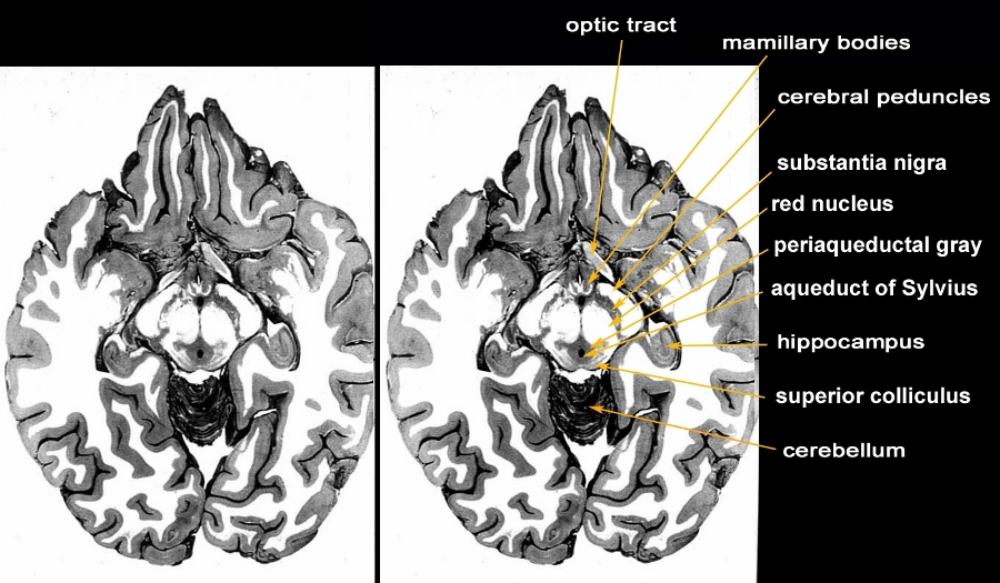
Axial Anatomic Specimen through the Midbrain Periaqueductal Gray Around the Aqueduct of Sylvius |
|
The anatomic section is an axial slice through the brain showing the structures of the midbrain. The most anterior structures of the midbrain are the cerebral peduncles followed by the substantia nigra, red nucleus, periaquaductal gray (PAG or central gray), aqueduct of Sylvius and finally the superior colliculus. Structures that are related to the midbrain anteriorly are the mamillary bodies and the optic tract. The hippocampus is positioned posterolaterally and the superior aspect of the cerebellum is seen posteriorly. Courtesy Department of Anatomy and Neurobiology at Boston University School of Medicine Dr. Jennifer Luebke , and Dr. Douglas Rosene 97140b03c.9 |
|
Whole Mount of the Midbrain in Axial Projection |
|
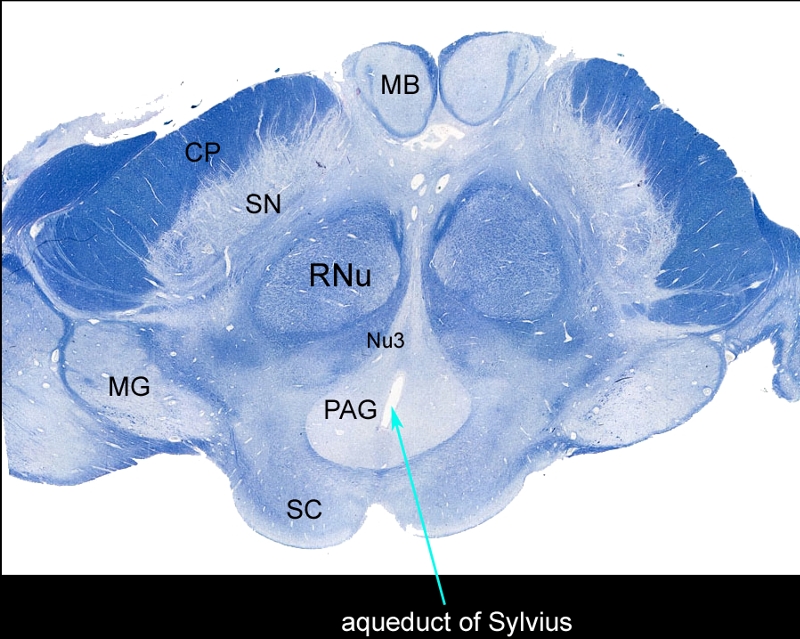
The mounted and stained midbrain in transverse plane illustrates the component structures. The anterior border of the midbrain incorporates the cerebral peduncles (CP), and the substantia nigra (SN). Between the substantia nigra and the aqueduct (teal arrow) is an area of the midbrain called the tegmentum (floor of the midbrain) Within the tegmentum other structures include red nuclii (RNu), oculomotor nucleus (Nu3), periaquaductal gray (PAG), and the aqueduct of Sylvius which is border forming between the tegmentum anteriorly and the tectum (roof) posteriorly. The posterior end of the midbrain is bordered by the colliculi in the tectum. Note also the paired mamillary bodies anteriorly (MB) and the medial geniculate body (MG) posterolaterally Courtesy Department of Anatomy and Neurobiology at Boston University School of Medicine Dr. Jennifer Luebke , and Dr. Douglas Rosene 98483L.8 |
Imaging
Coronal Plane
Aqueduct of Sylvius Finest of Channels in the Midbrain |
|
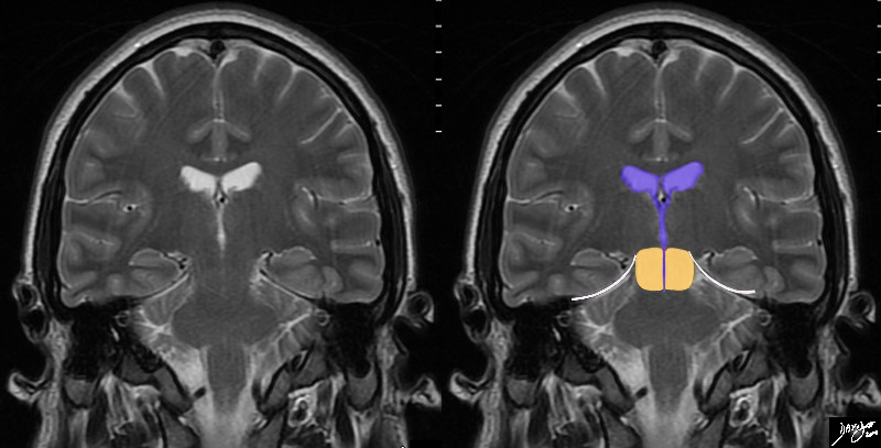
In this MRI image – the midbrain (orange) is seen centered aroubnd the ventriclar system. The aqueduct of Sylvius is the finest of channels that courses through the middle of the midbrain, and frequently is the key identifying feature of the midbrain. The temporal lobes rest of the tentorium (white curvilinear convex lines). Below the tenntorium is the midbrain and hind brain. Courtesy Ashley Davidoff MD copyright 2010 all rights reserved89721c03b01b.8s |
Sagittal Plane
 Aqueduct between the Tectum and Tegmentum Aqueduct between the Tectum and Tegmentum |
|
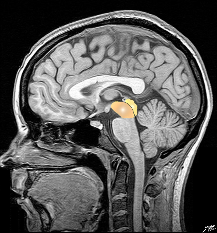
The midbrain consists of an anteroior tegmentum that incorporates the cerebral peduncles, (green and orange) divided from the posterior tectum consisting of the colliculi (purple) divided bythe cerebral aqueduct of Sylvius The Cerebral crura (green) are the most anteriorly placed, while the colliculi are the most posteriorly placed Courtesy Ashley Davidoff Md copyright 2010 all rights reserved 92141.3kb01bab02.8s |
Axial Plane
 Barely Visible Aqueduct of Sylvius Barely Visible Aqueduct of Sylvius
MRI T2 Weighted Image in the Axial Plane |
|
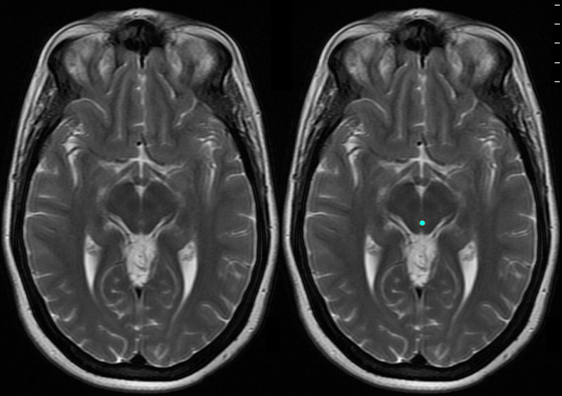
The axial T2 weighted MRI image through the midbrain shows the small but all important aqueduct of Sylvius (cerebral aqueduct) as it passes through the midbrain (Mickey Mouse appearance). Courtesy Ashley Davidoff MD copyright 2010 all rights reserved 94081c01.81s |
Applied Biology – Diseases
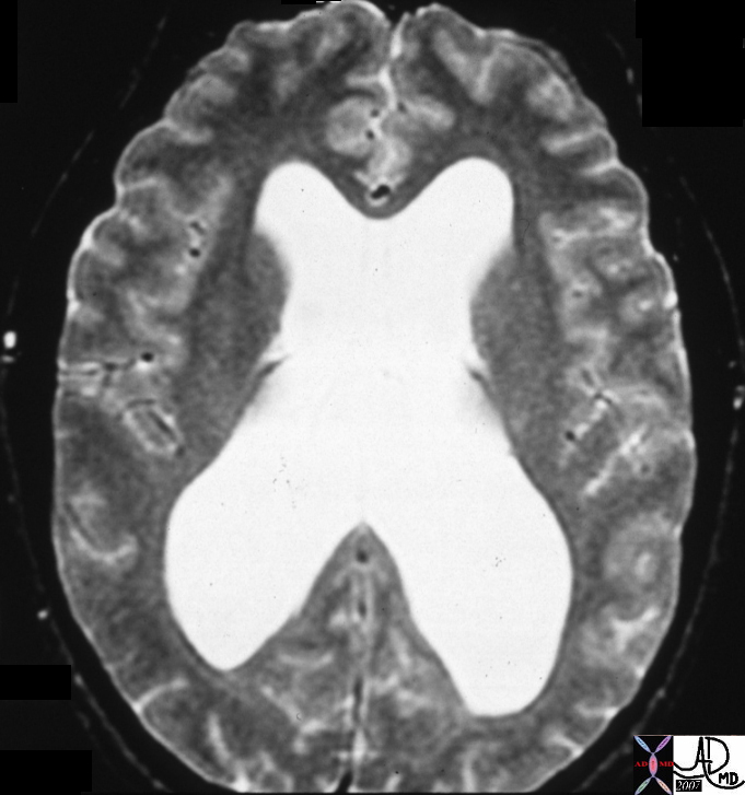 Aqueductal Stenosis Aqueductal Stenosis |
|
The T2 weighted MRI image in the axial plane shows very enlarged ventricles caused by aqueductal steniois which obstructs flow of the CSF through the aqueduct of Sylvius and results in hydrocephalus Courtesy James Donnelly MD 21644 |
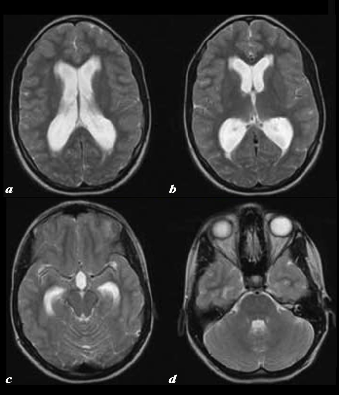 Aqueductal Stenosis Aqueductal Stenosis |
|
The T2 weighted MRI images show dilated lateral ventricles (a), frontal horns and occipital horns (b), temporal horns (c) and 4th ventricle (d) consistent with the known diagnosis of aqueductal stenosis CODE brain lateral ventricles frontal horns occipital horns atria body of temporal horns 4th ventricle aqueduct of Sylvius fx dilated enlarged distended dx hydrocephalus aqueductal stenosis Courtesy Philips Medical Systems 92408b.8 |
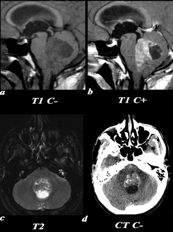 Medulloblastoma Impinging on the 4th ventricle Medulloblastoma Impinging on the 4th ventricle |
|
This 26 year old male presented with a three week history of progressive headache, nausea and vomiting. T1 pre (a) and post (b,): The exact origin of this mass can be difficult to ascertain given its large size. It clearly grows into the fourth ventricle, widening the lower portion of the Sylvian aqueduct. Note the smooth interface with the posterior aspect of the pons with associated mass effect verses the more irregular margin posteriorly where it arises from the cerebellum. T2 (c): The solid component of the medulloblastoma matches gray matter signal. Cystic or necrotic areas are demonstrated by higher T2 signal areas. CT(d): The unenhanced CT scan demonstrates a predominately hyperdense mass with punctuate calcifications in the region of the fourth ventricle . The low density area posteriorly is a cystic or necrotic component of the mass while the fourth ventricle is completely effaced. Image Courtesy Elisa Flower MD and Asim Mian MD 97668c01.81 |

