The Common Vein Copyright 2010
Definition
The atrium is part of the ventricular system of the brain and reoresents the part that connects the threee horns; frontal, occipital and temporal horns.
It usually contains the most columinous amount of choroid plexus which often calcifies in the region of the atrium.
|
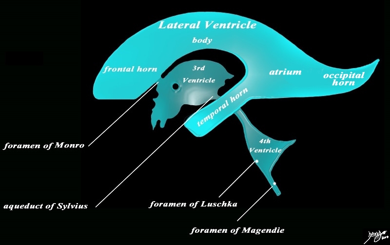
Sagittal View of the Ventricles |
|
The diagram in the sagittal projection reveals the horizontal portion called the lateral ventricle. It is a paired structure that houses the frontal horn, body, and the vertical portion which is composed of the 3rd ventricle, cerebral aqueduct and the 4th ventricle. The lateral ventricle consists of the frontal horn, body, occipital horn, atrium and the temporal horn The foramen of Monro connects the lateral ventricles with the third ventricle. The paired foramina of Luschka are sitiuated anteriorly in the 4th ventricle and they allow CSF to circulate in the subarachnoid spaces. The foramen of Magendie is a single structure and is situated posteriorly and it also enables CSF to enter the subarachnoid space. Courtesy Ashley Davidoff MD copyright 2010 all rights reserved 94459b10b02.82s |
|
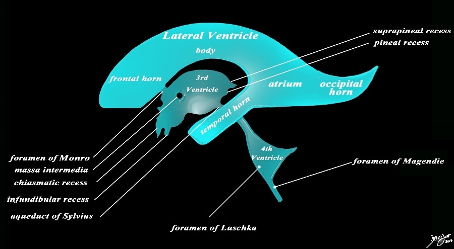
The Recesses |
|
The diagram in the sagittal projection reveals the horizontal portion called the lateral ventricle. It is a paired structure that houses the frontal horn, body, and the vertical portion which is composed of the 3rd ventricle, cerebral aqueduct and the 4th ventricle. The lateral ventricle consists of the frontal horn, body, occipital horn, atrium and the temporal horn The foramen of Monro connects the lateral ventricles with the third ventricle. The paired foramina of Luschka are sitiuated anteriorly in the 4th ventricle and they allow CSF to circulate in the subarachnoid spaces. The foramen of Magendie is a single structure and is situated posteriorly and it also enables CSF to enter the subarachnoid space. code brain ventricles lateral ventricles frontal horn body occipital horn atrium temporal horn 3rd ventricle 4th ventricle foramen of Monro formamen of Magendie Foramen of Luschka anatomy normal neuroanatomy diagram conceptual diagram structure principles Davidoff Art Courtesy Ashley Davidoff MD copyright 2010 all rights Courtesy Ashley Davidoff MD copyright 2010 all rights reserved 94459b10b02.82s |
|
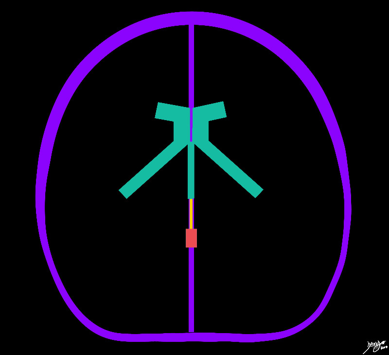
Atria – Part of the Forebrain – Midline and Medial Distribution of the Ventricles |
|
The distribution of the ventricular system is demonstrated in this diagram with the forebrain structures (green) including the lateral ventricles, frontal horns, atria, temporal horns, and third ventricle. The midbrain positioned aqueduct of Sylvius is seen in orange, while the hindbrained positioned 4th ventricle is overlaid in orange Courtesy Ashley DAvidoff MD copyright 2010 all rights reserved 93914b04d07.8s |
|
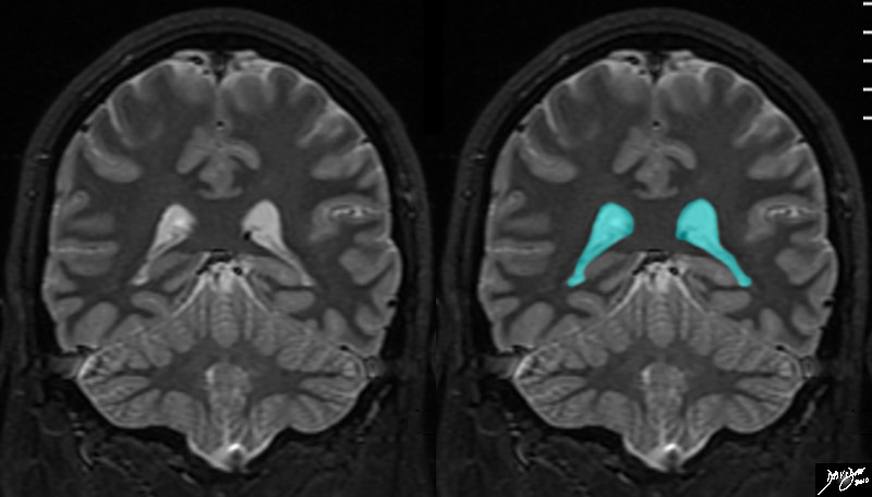
Temporal Horns Extending from the Atria |
|
The atria and temporal horns are demonstrated in this coronal MRI using a STIR sequence. The temporal horns are easily recognized as an inveted ‘V shaped structure starting roughly in the midline and coursing laterally and inferiorly. They start just anterior to the occipital lobes and end in the temporal lobes . Courtesy Ashley Davidoff MD copyright 2010 all rights reserved 89744c01.8s |
|
Choroid Plexus in the Temporal Horns |
|
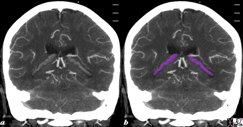
The reconstructed CTA scan in the coronal plane reveals the choroid plexus (purple) as fine knotty strands extending from the atrium to the temporal horns bilaterally. Image Courtesy Ashley Davidoff MD Copyright 2010 76532c.8s |
Applied Biology
|
Age related Atrophy Temporal Horns become Visible (d) |
|
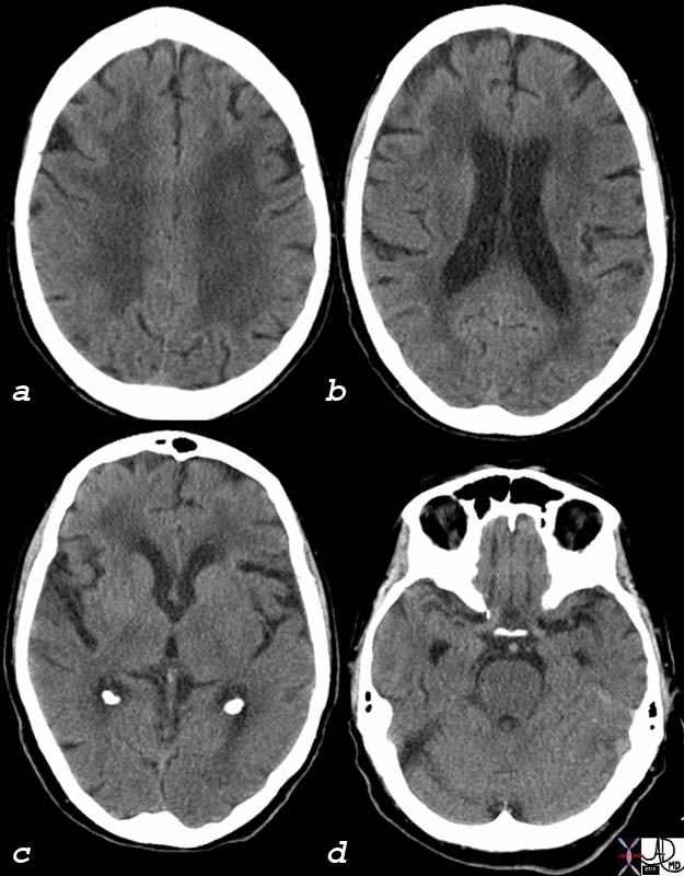
The CT scan of this 92 year old man reveals normal involutional change of the brain including perivntricular lucency(a)suggestive of microangiopathic change, mild dilatation of the ventricles (b) with deepening of the sulci and prominence of the gyri (abc) and ability to identify the temporal horns (d), all signs of brain atrophy. Note also the normal calcification of the choroid plexus in the atria (c) which is found very commonly even in young patients. Image Courtesy Ashley Davidoff MD 75932c01 |

