The Common vein Copyright 2010
Introduction

10 year old with Leukodystrophy |
|
This MRI is from a 10 year old male with known leukodystrophy. The findings on the MRI of the brain (a,b) are characterised by significant changes in the white matter of both frontal lobes. (green arrow), characterised by T2 and FLAIR hyperintensity These findings are consistent with leukodystrophy. Courtesy James Donnelly MD Copyright 2010 All rights reserved 23431c01 |

Adrenoleukodystrophy |
|
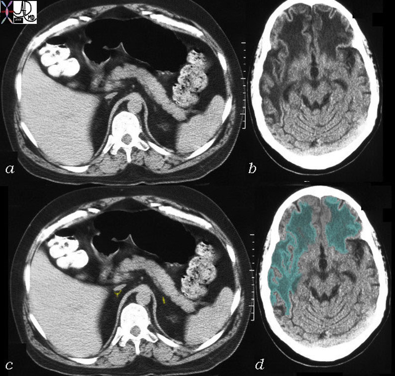
This CT is from a 26 year old male with known adrenoleukodystrophy. The findings on the CT of the brain (b,d) are characterised by significant changes in the white matter of both frontal lobes right parietal lobe and left parietal lobe to lesser extent. (green overlay) The adrenal glands (a,c) are barely visible but are overlaid in yellow in image (c). These findings are consistent with adrenoleukodystrophy. Courtesy Rebecca Schwartz MD Copyright 2010 All rights reserved 31464c04 |
