Oligodendroglioma
Copyright 2010
Definition
An oligodendroglioma is a type of glioma, that is, a tumor of the CNS that arises from glial cells. It makes up for about 20% to 30% of the gliomas, and occurs mostly in adults. Unlike the astrocytomas, they tend to be more present in the frontal and temporal lobes. On occasion, they may be multifocal however.
In the same way as other brain tumors, they present with focal cerebral dysfunction, which will vary and give a clue depending on location, and with generalized symptoms due to increased intracranial pressure.
Functionally, patients usually present with seizures, headaches, focal neurological deficits and personality changes. Seizures may be simple, complex partial, and even generalized.
These tumors are usually grossly soft, frequently with calcifications, hemorrhages, cysts.
Therefore, CT and MRI show calcifications; well demarcated tumors, without enhancement; the tumors are located near the cortical surface, with little or no edema. The tumors can be hemorrhagic (they are the most frequent intracranial tumor that bleeds) or be cystic.
More than half of the oligodendrogliomas are sensitive to PCV chemotherapy and radiation. Complete surgical resection is many times chosen however, as it also improves survival.

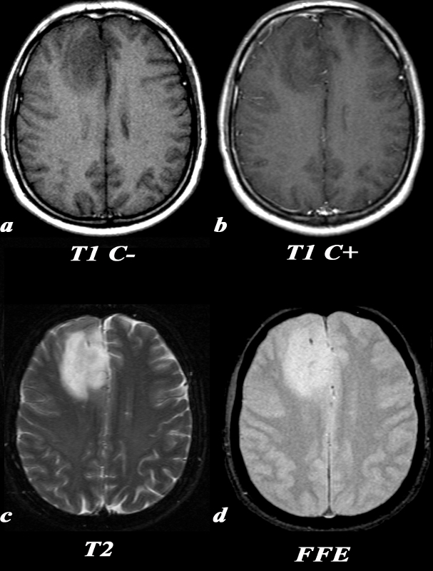
Frontal Oligodendroglioma with Calcification |
|
This 35 year old male presented with new onset of seizure. T1 C- (a): On this T1 weighted image, there is a hypointense region in the right frontal lobe with blurring of the normally clear gray-white matter distinction. T1 post C+ (b): No definite areas of enhancement were identified in this mass which is occasionally the case in oligodendrogliomas. T2 (c): On T2 images, this lesion is more clearly defined as a region of increased signal. There is expansion of the involved gyri, as the adjacent sulci are not as clearly seen as they are in the same region on the contralateral side. Note the relative lack of surrounding edema around the lesion which would have the appearance of increased signal in the adjacent white matter sparing the gray matter. FFE(d): This gradient echo MRI sequence is tailored for picking up substances which alter the local magnetic field, or paramagnetic substances, which will appear hypointense. This “blooming” can be seen in areas of calcification, as in this case. Calcification is seen in a majority of oligodendrogliomas. Image Courtesy Lawrence Chin MD 97673c.8 |
