The Common Vein Copyright 2010
Introduction
Abscess

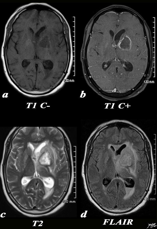
Rim Enhancement with Gadolinium |
|
The basal ganglia in the region of the caudate nucleus and globus pallidus are shown in axial projection in this 60 year old female who presents with neurological deficit and a fever. In the first image the focal ill defined mass with mild mass effect is shown in axial projection on a T1 weighted image without contrast (a). The second image with gadolinium shows rim enhancement with mass effect and obstruction of the frontal horn as seen by asymmetric dilatation (b). The third T2 weighted image (c) shows the fluid nature of the cavity and the surrounding edema, mass effect, and accumulation of fluid in the dependant portion of the occipital horn. The fourth image is a FLAIR image and also shows th extent of the edema in the brain The patient had a fever and the constellation of findings were consistent with an abscess of the basal ganglia on the left. Courtesy Ashley Davidoff MD Copyright 2010 All rights reserved 89054c.8s |
Tumors

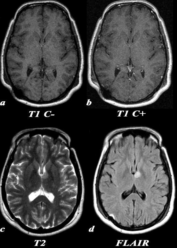
Low Grade Astrocytoma not Visualized on Contrast Enhanced Study |
|
A 31 year old female presented with transient episodes of numbness and tingling in her right face, arm and leg. MRI: T1 C- (a) No definite signal abnormality is identified in the left thalamic low grade glioma. T1 post (T1 C+ b) : Post contrast images demonstrate no enhancement in the region of the known mass. This is typical of a low grade glioma. T2 (c): This T2 weighted image shows a unilateral well circumscribed area of increased signal in the anterior left thalamus. There is no significant mass effect on the adjacent structures. FLAIR (d): This FLAIR image from her MRI demonstrates (with slightly better clarity than the T2 weighted image) a unilateral well circumscribed area of increased signal in the anterior left thalamus. There is no significant mass effect on the adjacent structures. Image Courtesy Elisa Flower MD and Asim Mian MD 97664c.8 |
